6CDJ | pdb_00006cdj

HIV-1 wild type protease with GRL-03314A, 6-5-5-ring fused umbrella-like tetrahydropyranofuran as the P2-ligand, a cyclopropylaminobenzothiazole as the P2'-ligand and 3,5-difluorophenylmethyl as the P1-ligand


Experimental Data Snapshot

  • Method: X-RAY DIFFRACTION
  • Resolution: 1.13 Å
  • R-Value Free: 
    0.173 (Depositor), 0.174 (DCC) 
  • R-Value Work: 
    0.142 (Depositor), 0.150 (DCC) 
  • R-Value Observed: 
    0.144 (Depositor) 

Starting Model: experimental
View more details

wwPDB Validation 3D Report Full Report

Validation slider image for 6CDJ

Ligand Structure Quality Assessment 


This is version 1.4 of the entry. See complete history

Literature

Design and Synthesis of Highly Potent HIV-1 Protease Inhibitors Containing Tricyclic Fused Ring Systems as Novel P2 Ligands: Structure-Activity Studies, Biological and X-ray Structural Analysis.

Ghosh, A.K.R Nyalapatla, P.Kovela, S.Rao, K.V.Brindisi, M.Osswald, H.L.Amano, M.Aoki, M.Agniswamy, J.Wang, Y.F.Weber, I.T.Mitsuya, H.

(2018) J Med Chem 61: 4561-4577

  • DOI: https://doi.org/10.1021/acs.jmedchem.8b00298
  • Primary Citation Related Structures: 
    6CDJ, 6CDL

  • PubMed Abstract: 

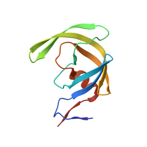
    The design, synthesis, and biological evaluation of a new class of HIV-1 protease inhibitors containing stereochemically defined fused tricyclic polyethers as the P2 ligands and a variety of sulfonamide derivatives as the P2' ligands are described. A number of ring sizes and various substituent effects were investigated to enhance the ligand-backbone interactions in the protease active site. Inhibitors 5c and 5d containing this unprecedented fused 6-5-5 ring system as the P2 ligand, an aminobenzothiazole as the P2' ligand, and a difluorophenylmethyl as the P1 ligand exhibited exceptional enzyme inhibitory potency and maintained excellent antiviral activity against a panel of highly multidrug-resistant HIV-1 variants. The umbrella-like P2 ligand for these inhibitors has been synthesized efficiently in an optically active form using a Pauson-Khand cyclization reaction as the key step. The racemic alcohols were resolved efficiently using a lipase catalyzed enzymatic resolution. Two high resolution X-ray structures of inhibitor-bound HIV-1 protease revealed extensive interactions with the backbone atoms of HIV-1 protease and provided molecular insight into the binding properties of these new inhibitors.


  • Organizational Affiliation
    • Department of Chemistry and Department of Medicinal Chemistry , Purdue University , 560 Oval Drive , West Lafayette , Indiana 47907 , United States.

Macromolecule Content 

  • Total Structure Weight: 22.54 kDa 
  • Atom Count: 2,023 
  • Modeled Residue Count: 198 
  • Deposited Residue Count: 198 
  • Unique protein chains: 1

Macromolecules

Find similar proteins by:|  3D Structure
Entity ID: 1
MoleculeChains  Sequence LengthOrganismDetailsImage
Protease
A, B
99Human immunodeficiency virus 1Mutation(s): 5 
Gene Names: pol
UniProt
Find proteins for Q5RZ08 (Human immunodeficiency virus type 1)
Explore Q5RZ08 
Go to UniProtKB:  Q5RZ08
Entity Groups
Sequence Clusters30% Identity50% Identity70% Identity90% Identity95% Identity100% Identity
UniProt GroupQ5RZ08
Sequence Annotations
Expand
Reference Sequence

Small Molecules

Ligands 5 Unique
IDChains Name / Formula / InChI Key2D Diagram3D Interactions
GR8

Query on GR8


Download:Ideal Coordinates CCD File
C [auth A](2aS,4R,4aS,7aS,7bS)-octahydro-2H-1,7-dioxacyclopenta[cd]inden-4-yl [(2S,3R)-4-[{[2-(cyclopropylamino)-1,3-benzothiazol-6-yl]sulfonyl}(2-methylpropyl)amino]-1-(3,5-difluorophenyl)-3-hydroxybutan-2-yl]carbamate
C34 H42 F2 N4 O7 S2
NIZKZMLJMFCQSW-FQOBIZQLSA-N
GOL

Query on GOL


Download:Ideal Coordinates CCD File
F [auth A]GLYCEROL
C3 H8 O3
PEDCQBHIVMGVHV-UHFFFAOYSA-N
ACT

Query on ACT


Download:Ideal Coordinates CCD File
I [auth B],
J [auth B]
ACETATE ION
C2 H3 O2
QTBSBXVTEAMEQO-UHFFFAOYSA-M
CL

Query on CL


Download:Ideal Coordinates CCD File
E [auth A],
G [auth B],
H [auth B]
CHLORIDE ION
Cl
VEXZGXHMUGYJMC-UHFFFAOYSA-M
NA

Query on NA


Download:Ideal Coordinates CCD File
D [auth A]SODIUM ION
Na
FKNQFGJONOIPTF-UHFFFAOYSA-N

Experimental Data & Validation

Experimental Data

  • Method: X-RAY DIFFRACTION
  • Resolution: 1.13 Å
  • R-Value Free:  0.173 (Depositor), 0.174 (DCC) 
  • R-Value Work:  0.142 (Depositor), 0.150 (DCC) 
  • R-Value Observed: 0.144 (Depositor) 
Space Group: P 21 21 2
Unit Cell:
Length ( Å )Angle ( ˚ )
a = 58.959α = 90
b = 86.077β = 90
c = 46.157γ = 90
Software Package:
Software NamePurpose
HKL-2000data reduction
HKL-2000data scaling
PHASERphasing
SHELXL-97refinement

Structure Validation

View Full Validation Report


Ligand Structure Quality Assessment 


Entry History 

& Funding Information

Deposition Data


Funding OrganizationLocationGrant Number
National Institutes of Health/National Institute of General Medical Sciences (NIH/NIGMS)United StatesGM53386
National Institutes of Health/National Institute of General Medical Sciences (NIH/NIGMS)United StatesGM62920
National Institutes of Health/National Cancer Institute (NIH/NCI)United Statesthe Intramural Research Program of the Center for Cancer Research
Ministry of Education, Culture, Sports, Science and Technology (Japan)Japana Grant-in-Aid for Scientific Research (Priority Areas)
Ministry of Health, Welfare, and LaborJapana Grant for Promotion of AIDS Research
Ministry of Education, Culture, Sports, Science and Technology (Japan)Japanthe Grant to the Cooperative Research Project on Clinical and Epidemiological Studies of Emerging and Reemerging Infectious Diseases (Renkei Jigyo)

Revision History  (Full details and data files)

  • Version 1.0: 2018-05-30
    Type: Initial release
  • Version 1.1: 2018-06-06
    Changes: Data collection, Database references
  • Version 1.2: 2019-02-20
    Changes: Author supporting evidence, Data collection
  • Version 1.3: 2019-12-04
    Changes: Author supporting evidence
  • Version 1.4: 2023-10-04
    Changes: Data collection, Database references, Derived calculations, Refinement description