6CDZ

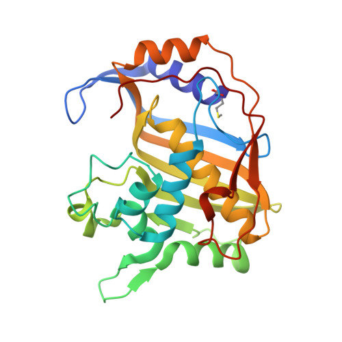
E. coli thymidylate synthase mutant I264Am


Experimental Data Snapshot

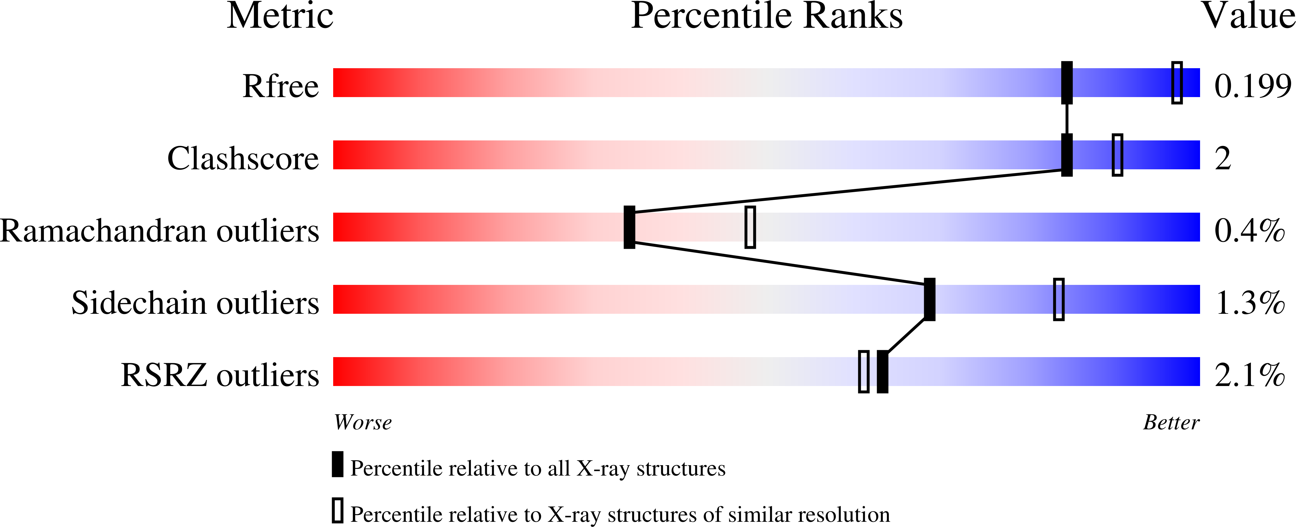
  • Method: X-RAY DIFFRACTION
  • Resolution: 2.40 Å
  • R-Value Free: 0.229 
  • R-Value Work: 0.180 
  • R-Value Observed: 0.183 

Starting Model: experimental
View more details

wwPDB Validation   3D Report Full Report


Ligand Structure Quality Assessment 


This is version 1.4 of the entry. See complete history


Literature

A Single Mutation Traps a Half-Sites Reactive Enzyme in Midstream, Explaining Asymmetry in Hydride Transfer.

Finer-Moore, J.S.Lee, T.T.Stroud, R.M.

(2018) Biochemistry 57: 2786-2795

  • DOI: https://doi.org/10.1021/acs.biochem.8b00176
  • Primary Citation of Related Structures:  
    6CDZ

  • PubMed Abstract: 

    In Escherichia coli thymidylate synthase (EcTS), rate-determining hydride transfer from the cofactor 5,10-methylene-5,6,7,8-tetrahydrofolate to the intermediate 5-methylene-2'-deoxyuridine 5'-monophosphate occurs by hydrogen tunneling, requiring precise alignment of reactants and a closed binding cavity, sealed by the C-terminal carboxyl group. Mutations that destabilize the closed conformation of the binding cavity allow small molecules such as β-mercaptoethanol (β-ME) to enter the active site and compete with hydride for addition to the 5-methylene group of the intermediate. The C-terminal deletion mutant of EcTS produced the β-ME adduct in proportions that varied dramatically with cofactor concentration, from 50% at low cofactor concentrations to 0% at saturating cofactor conditions, suggesting communication between active sites. We report the 2.4 Å X-ray structure of the C-terminal deletion mutant of E. coli TS in complex with a substrate and a cofactor analogue, CB3717. The structure is asymmetric, with reactants aligned in a manner consistent with hydride transfer in only one active site. In the second site, CB3717 has shifted to a site where the normal cofactor would be unlikely to form 5-methylene-2'-deoxyuridine 5'-monophosphate, consistent with no formation of the β-ME adduct. The structure shows how the binding of the cofactor at one site triggers hydride transfer and borrows needed stabilization from substrate binding at the second site. It indicates pathways through the dimer interface that contribute to allostery relevant to half-sites reactivity.


  • Organizational Affiliation

    Department of Biochemistry and Biophysics , University of California , San Francisco , California 94143-2240 , United States.


Macromolecules
Find similar proteins by:  (by identity cutoff)  |  3D Structure
Entity ID: 1
MoleculeChains Sequence LengthOrganismDetailsImage
Thymidylate synthase
A, B
263Escherichia coli K-12Mutation(s): 0 
Gene Names: thyAb2827JW2795
EC: 2.1.1.45
UniProt
Find proteins for P0A884 (Escherichia coli (strain K12))
Explore P0A884 
Go to UniProtKB:  P0A884
Entity Groups  
Sequence Clusters30% Identity50% Identity70% Identity90% Identity95% Identity100% Identity
UniProt GroupP0A884
Sequence Annotations
Expand
  • Reference Sequence
Experimental Data & Validation

Experimental Data

  • Method: X-RAY DIFFRACTION
  • Resolution: 2.40 Å
  • R-Value Free: 0.229 
  • R-Value Work: 0.180 
  • R-Value Observed: 0.183 
  • Space Group: C 1 2 1
Unit Cell:
Length ( Å )Angle ( ˚ )
a = 67.658α = 90
b = 80.451β = 103.5
c = 94.08γ = 90
Software Package:
Software NamePurpose
PHENIXrefinement
DENZOdata reduction
SCALEPACKdata scaling
AMoREphasing

Structure Validation

View Full Validation Report


Ligand Structure Quality Assessment 


Entry History & Funding Information

Deposition Data


Funding OrganizationLocationGrant Number
National Institutes of Health/National Cancer Institute (NIH/NCI)United StatesCA-41323
National Institutes of Health/National Cancer Institute (NIH/NCI)United StatesCA63081
National Institutes of Health/National Institute of General Medical Sciences (NIH/NIGMS)United StatesGM024485

Revision History  (Full details and data files)

  • Version 1.0: 2018-05-16
    Type: Initial release
  • Version 1.1: 2018-05-23
    Changes: Data collection, Database references
  • Version 1.2: 2019-12-04
    Changes: Author supporting evidence
  • Version 1.3: 2023-10-04
    Changes: Data collection, Database references, Refinement description
  • Version 1.4: 2024-10-16
    Changes: Structure summary