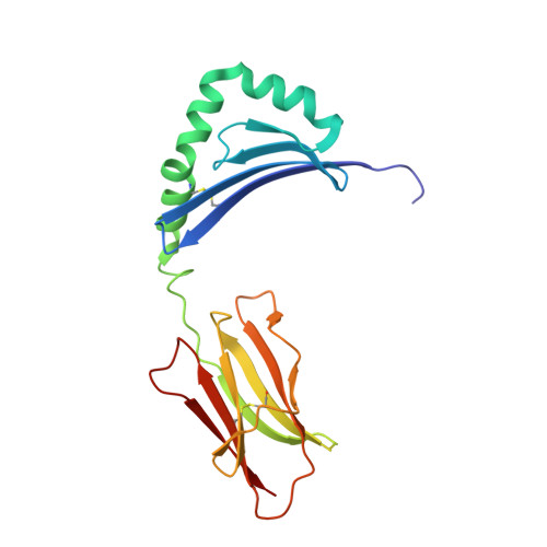
6CQJ

Crystal structure of DR1 presenting the RQ13 peptide


Experimental Data Snapshot

  • Method: X-RAY DIFFRACTION
  • Resolution: 2.75 Å
  • R-Value Free: 0.249 
  • R-Value Work: 0.197 
  • R-Value Observed: 0.200 

Starting Model: experimental
View more details

wwPDB Validation   3D Report Full Report


Ligand Structure Quality Assessment 


This is version 2.2 of the entry. See complete history


Literature

CD4+T cell-mediated HLA class II cross-restriction in HIV controllers.

Galperin, M.Farenc, C.Mukhopadhyay, M.Jayasinghe, D.Decroos, A.Benati, D.Tan, L.L.Ciacchi, L.Reid, H.H.Rossjohn, J.Chakrabarti, L.A.Gras, S.

(2018) Sci Immunol 3

  • DOI: https://doi.org/10.1126/sciimmunol.aat0687
  • Primary Citation of Related Structures:  
    6CPH, 6CPL, 6CPN, 6CPO, 6CQJ, 6CQL, 6CQN, 6CQQ, 6CQR

  • PubMed Abstract: 

    Rare individuals, termed HIV controllers, spontaneously control HIV infection by mounting efficient T cell responses against the virus. Protective CD4 + T cell responses from HIV controllers involve high-affinity public T cell receptors (TCRs) recognizing an immunodominant capsid epitope (Gag293) presented by a remarkably broad array of human leukocyte antigen (HLA) class II molecules. Here, we determine the structures of a prototypical public TCR bound to HLA-DR1, HLA-DR11, and HLA-DR15 molecules presenting the Gag293 epitope. TCR recognition was driven by contacts with the Gag293 epitope, a feature that underpinned the extensive HLA cross-restriction. These high-affinity TCRs promoted mature immunological synapse formation and cytotoxic capacity in both CD4 + and CD8 + T cells. The public TCRs suppressed HIV replication in multiple genetic backgrounds ex vivo, emphasizing the functional advantage conferred by broad HLA class II cross-restriction.


  • Organizational Affiliation

    Pasteur Institute, Viral Pathogenesis Unit, Paris, France.


Macromolecules
Find similar proteins by:  (by identity cutoff)  |  3D Structure
Entity ID: 1
MoleculeChains Sequence LengthOrganismDetailsImage
HLA class II histocompatibility antigen, DR alpha chain
A, D, G
182Homo sapiensMutation(s): 0 
Gene Names: HLA-DRAHLA-DRA1
UniProt & NIH Common Fund Data Resources
Find proteins for P01903 (Homo sapiens)
Explore P01903 
Go to UniProtKB:  P01903
PHAROS:  P01903
GTEx:  ENSG00000204287 
Entity Groups  
Sequence Clusters30% Identity50% Identity70% Identity90% Identity95% Identity100% Identity
UniProt GroupP01903
Glycosylation
Glycosylation Sites: 1Go to GlyGen: P01903-1
Sequence Annotations
Expand
  • Reference Sequence
Find similar proteins by:  (by identity cutoff)  |  3D Structure
Entity ID: 2
MoleculeChains Sequence LengthOrganismDetailsImage
HLA class II histocompatibility antigen, DRB1-1 beta chain
B, E, H
189Homo sapiensMutation(s): 0 
Gene Names: HLA-DRB1
UniProt & NIH Common Fund Data Resources
Find proteins for P01911 (Homo sapiens)
Explore P01911 
Go to UniProtKB:  P01911
PHAROS:  P01911
GTEx:  ENSG00000196126 
Entity Groups  
Sequence Clusters30% Identity50% Identity70% Identity90% Identity95% Identity100% Identity
UniProt GroupP01911
Sequence Annotations
Expand
  • Reference Sequence

Find similar proteins by:  Sequence   |   3D Structure  

Entity ID: 3
MoleculeChains Sequence LengthOrganismDetailsImage
Peptide from Capsid protein p24
C, F, I
13HIV-1 M:B_HXB2RMutation(s): 0 
UniProt
Find proteins for P04585 (Human immunodeficiency virus type 1 group M subtype B (isolate HXB2))
Explore P04585 
Go to UniProtKB:  P04585
Entity Groups  
Sequence Clusters30% Identity50% Identity70% Identity90% Identity95% Identity100% Identity
UniProt GroupP04585
Sequence Annotations
Expand
  • Reference Sequence
Oligosaccharides

Help

Entity ID: 4
MoleculeChains Length2D Diagram Glycosylation3D Interactions
2-acetamido-2-deoxy-beta-D-glucopyranose-(1-4)-2-acetamido-2-deoxy-beta-D-glucopyranose-(1-4)-2-acetamido-2-deoxy-beta-D-glucopyranose
J
3N-Glycosylation
Glycosylation Resources
GlyTouCan:  G47362BJ
GlyCosmos:  G47362BJ
GlyGen:  G47362BJ
Small Molecules
Ligands 3 Unique
IDChains Name / Formula / InChI Key2D Diagram3D Interactions
NAG
Query on NAG

Download Ideal Coordinates CCD File 
L [auth A],
O [auth D]
2-acetamido-2-deoxy-beta-D-glucopyranose
C8 H15 N O6
OVRNDRQMDRJTHS-FMDGEEDCSA-N
PEG
Query on PEG

Download Ideal Coordinates CCD File 
K [auth A],
M [auth B],
P [auth E],
Q [auth G]
DI(HYDROXYETHYL)ETHER
C4 H10 O3
MTHSVFCYNBDYFN-UHFFFAOYSA-N
NA
Query on NA

Download Ideal Coordinates CCD File 
N [auth D],
R [auth G]
SODIUM ION
Na
FKNQFGJONOIPTF-UHFFFAOYSA-N
Biologically Interesting Molecules (External Reference) 1 Unique
Experimental Data & Validation

Experimental Data

  • Method: X-RAY DIFFRACTION
  • Resolution: 2.75 Å
  • R-Value Free: 0.249 
  • R-Value Work: 0.197 
  • R-Value Observed: 0.200 
  • Space Group: P 1
Unit Cell:
Length ( Å )Angle ( ˚ )
a = 68.684α = 61.56
b = 82.017β = 88.2
c = 83.05γ = 86.47
Software Package:
Software NamePurpose
BUSTERrefinement
XDSdata reduction
Aimlessdata scaling

Structure Validation

View Full Validation Report


Ligand Structure Quality Assessment 


Entry History 

Deposition Data

Revision History  (Full details and data files)

  • Version 1.0: 2018-06-06
    Type: Initial release
  • Version 1.1: 2018-06-27
    Changes: Data collection, Database references, Structure summary
  • Version 2.0: 2020-07-29
    Type: Remediation
    Reason: Carbohydrate remediation
    Changes: Atomic model, Data collection, Derived calculations, Structure summary
  • Version 2.1: 2023-10-04
    Changes: Data collection, Database references, Refinement description, Structure summary
  • Version 2.2: 2024-10-23
    Changes: Structure summary