6CWY

Crystal structure of SUMO E1 in complex with an allosteric inhibitor


Experimental Data Snapshot

  • Method: X-RAY DIFFRACTION
  • Resolution: 2.46 Å
  • R-Value Free: 
    0.237 (Depositor), 0.240 (DCC) 
  • R-Value Work: 
    0.197 (Depositor), 0.200 (DCC) 
  • R-Value Observed: 
    0.199 (Depositor) 

Starting Model: experimental
View more details

wwPDB Validation   3D Report Full Report


Ligand Structure Quality Assessment 

Created with Raphaël 2.3.0Worse 01 BetterLigand structure goodness of fit to experimental dataBest fitted FHJClick on this verticalbar to view details

This is version 1.3 of the entry. See complete history


Literature

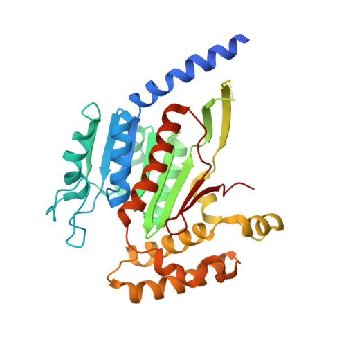
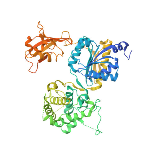
Molecular mechanism of a covalent allosteric inhibitor of SUMO E1 activating enzyme.

Lv, Z.Yuan, L.Atkison, J.H.Williams, K.M.Vega, R.Sessions, E.H.Divlianska, D.B.Davies, C.Chen, Y.Olsen, S.K.

(2018) Nat Commun 9: 5145-5145

  • DOI: https://doi.org/10.1038/s41467-018-07015-1
  • Primary Citation of Related Structures:  
    6CWY, 6CWZ

  • PubMed Abstract: 

    E1 enzymes activate ubiquitin (Ub) and ubiquitin-like modifiers (Ubls) in the first step of Ub/Ubl conjugation cascades and represent potential targets for therapeutic intervention in cancer and other life-threatening diseases. Here, we report the crystal structure of the E1 enzyme for the Ubl SUMO in complex with a recently discovered and highly specific covalent allosteric inhibitor (COH000). The structure reveals that COH000 targets a cryptic pocket distinct from the active site that is completely buried in all previous SUMO E1 structures and that COH000 binding to SUMO E1 is accompanied by a network of structural changes that altogether lock the enzyme in a previously unobserved inactive conformation. These structural changes include disassembly of the active site and a 180° rotation of the catalytic cysteine-containing SCCH domain, relative to conformational snapshots of SUMO E1 poised to catalyze adenylation. Altogether, our study provides a molecular basis for the inhibitory mechanism of COH000 and its SUMO E1 specificity, and also establishes a framework for potential development of molecules targeting E1 enzymes for other Ubls at a cryptic allosteric site.


  • Organizational Affiliation

    Department of Biochemistry & Molecular Biology and Hollings Cancer Center, Medical University of South Carolina, Charleston, 29425, SC, USA.


Macromolecules
Find similar proteins by:  (by identity cutoff)  |  3D Structure
Entity ID: 1
MoleculeChains Sequence LengthOrganismDetailsImage
SUMO-activating enzyme subunit 1A [auth C]346Homo sapiensMutation(s): 0 
Gene Names: SAE1AOS1SUA1UBLE1A
UniProt & NIH Common Fund Data Resources
Find proteins for Q9UBE0 (Homo sapiens)
Explore Q9UBE0 
Go to UniProtKB:  Q9UBE0
PHAROS:  Q9UBE0
GTEx:  ENSG00000142230 
Entity Groups  
Sequence Clusters30% Identity50% Identity70% Identity90% Identity95% Identity100% Identity
UniProt GroupQ9UBE0
Sequence Annotations
Expand
  • Reference Sequence
Find similar proteins by:  (by identity cutoff)  |  3D Structure
Entity ID: 2
MoleculeChains Sequence LengthOrganismDetailsImage
SUMO-activating enzyme subunit 2B [auth D]660Homo sapiensMutation(s): 0 
Gene Names: UBA2SAE2UBLE1BHRIHFB2115
EC: 2.3.2
UniProt & NIH Common Fund Data Resources
Find proteins for Q9UBT2 (Homo sapiens)
Explore Q9UBT2 
Go to UniProtKB:  Q9UBT2
PHAROS:  Q9UBT2
GTEx:  ENSG00000126261 
Entity Groups  
Sequence Clusters30% Identity50% Identity70% Identity90% Identity95% Identity100% Identity
UniProt GroupQ9UBT2
Sequence Annotations
Expand
  • Reference Sequence
Small Molecules
Ligands 5 Unique
IDChains Name / Formula / InChI Key2D Diagram3D Interactions
FHJ
Query on FHJ

Download Ideal Coordinates CCD File 
K [auth D]dimethyl (1S,2S,3R,4R)-1-[(1S)-2-(4-methylphenyl)-1-(phenylamino)ethyl]-7-oxabicyclo[2.2.1]hept-5-ene-2,3-dicarboxylate
C25 H27 N O5
AOCBOTNEIQNHIY-OAKYZLIPSA-N
SO4
Query on SO4

Download Ideal Coordinates CCD File 
G [auth D],
H [auth D],
I [auth D],
J [auth D]
SULFATE ION
O4 S
QAOWNCQODCNURD-UHFFFAOYSA-L
GOL
Query on GOL

Download Ideal Coordinates CCD File 
C,
D [auth C]
GLYCEROL
C3 H8 O3
PEDCQBHIVMGVHV-UHFFFAOYSA-N
ZN
Query on ZN

Download Ideal Coordinates CCD File 
E [auth D]ZINC ION
Zn
PTFCDOFLOPIGGS-UHFFFAOYSA-N
MG
Query on MG

Download Ideal Coordinates CCD File 
F [auth D]MAGNESIUM ION
Mg
JLVVSXFLKOJNIY-UHFFFAOYSA-N
Experimental Data & Validation

Experimental Data

  • Method: X-RAY DIFFRACTION
  • Resolution: 2.46 Å
  • R-Value Free:  0.237 (Depositor), 0.240 (DCC) 
  • R-Value Work:  0.197 (Depositor), 0.200 (DCC) 
  • R-Value Observed: 0.199 (Depositor) 
Space Group: P 21 21 21
Unit Cell:
Length ( Å )Angle ( ˚ )
a = 56.055α = 90
b = 116.046β = 90
c = 174.143γ = 90
Software Package:
Software NamePurpose
PHENIXrefinement
HKL-2000data reduction
HKL-2000data scaling
PDB_EXTRACTdata extraction
PHASERphasing

Structure Validation

View Full Validation Report


Ligand Structure Quality Assessment 

Created with Raphaël 2.3.0Worse 01 BetterLigand structure goodness of fit to experimental dataBest fitted FHJClick on this verticalbar to view details

Entry History & Funding Information

Deposition Data


Funding OrganizationLocationGrant Number
National Institutes of Health/National Institute of General Medical Sciences (NIH/NIGMS)United StatesGM115568-02

Revision History  (Full details and data files)

  • Version 1.0: 2019-01-16
    Type: Initial release
  • Version 1.1: 2020-01-01
    Changes: Author supporting evidence
  • Version 1.2: 2023-10-04
    Changes: Data collection, Database references, Refinement description
  • Version 1.3: 2024-10-23
    Changes: Structure summary