6D42

Crystal structure of the KCa3.1 C-terminal four-helix bundle (with copper)

  • Classification: MEMBRANE PROTEIN
  • Organism(s): Homo sapiens
  • Expression System: Escherichia coli
  • Mutation(s): No 

  • Deposited: 2018-04-17 Released: 2018-07-11 
  • Deposition Author(s): Hubbard, S.R., Ji, T.
  • Funding Organization(s): National Institutes of Health/National Institute Of Allergy and Infectious Diseases (NIH/NIAID)

Experimental Data Snapshot

  • Method: X-RAY DIFFRACTION
  • Resolution: 1.75 Å
  • R-Value Free: 0.323 
  • R-Value Work: 0.252 
  • R-Value Observed: 0.259 

wwPDB Validation   3D Report Full Report


This is version 1.1 of the entry. See complete history


Literature

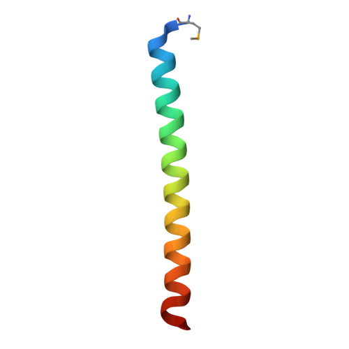
Crystal structure of the C-terminal four-helix bundle of the potassium channel KCa3.1.

Ji, T.Corbalan-Garcia, S.Hubbard, S.R.

(2018) PLoS One 13: e0199942-e0199942

  • DOI: https://doi.org/10.1371/journal.pone.0199942
  • Primary Citation of Related Structures:  
    6D42

  • PubMed Abstract: 

    KCa3.1 (also known as SK4 or IK1) is a mammalian intermediate-conductance potassium channel that plays a critical role in the activation of T cells, B cells, and mast cells, effluxing potassium ions to maintain a negative membrane potential for influxing calcium ions. KCa3.1 shares primary sequence similarity with three other (low-conductance) potassium channels: KCa2.1, KCa2.2, and KCa2.3 (also known as SK1-3). These four homotetrameric channels bind calmodulin (CaM) in the cytoplasmic region, and calcium binding to CaM triggers channel activation. Unique to KCa3.1, activation also requires phosphorylation of a single histidine residue, His358, in the cytoplasmic region, which relieves copper-mediated inhibition of the channel. Near the cytoplasmic C-terminus of KCa3.1 (and KCa2.1-2.3), secondary-structure analysis predicts the presence of a coiled-coil/heptad repeat. Here, we report the crystal structure of the C-terminal coiled-coil region of KCa3.1, which forms a parallel four-helix bundle, consistent with the tetrameric nature of the channel. Interestingly, the four copies of a histidine residue, His389, in an 'a' position within the heptad repeat, are observed to bind a copper ion along the four-fold axis of the bundle. These results suggest that His358, the inhibitory histidine in KCa3.1, might coordinate a copper ion through a similar binding mode.


  • Organizational Affiliation

    Kimmel Center for Biology and Medicine of the Skirball Institute, Department of Biochemistry and Molecular Pharmacology, New York University School of Medicine, New York, NY, United States of America.


Macromolecules
Find similar proteins by:  (by identity cutoff)  |  3D Structure
Entity ID: 1
MoleculeChains Sequence LengthOrganismDetailsImage
Intermediate conductance calcium-activated potassium channel protein 4
A, B
42Homo sapiensMutation(s): 0 
Gene Names: KCNN4IK1IKCA1KCA4SK4
UniProt & NIH Common Fund Data Resources
Find proteins for O15554 (Homo sapiens)
Explore O15554 
Go to UniProtKB:  O15554
PHAROS:  O15554
GTEx:  ENSG00000104783 
Entity Groups  
Sequence Clusters30% Identity50% Identity70% Identity90% Identity95% Identity100% Identity
UniProt GroupO15554
Sequence Annotations
Expand
  • Reference Sequence
Small Molecules
Modified Residues  1 Unique
IDChains TypeFormula2D DiagramParent
MSE
Query on MSE
A, B
L-PEPTIDE LINKINGC5 H11 N O2 SeMET
Experimental Data & Validation

Experimental Data

  • Method: X-RAY DIFFRACTION
  • Resolution: 1.75 Å
  • R-Value Free: 0.323 
  • R-Value Work: 0.252 
  • R-Value Observed: 0.259 
  • Space Group: P 4 21 2
Unit Cell:
Length ( Å )Angle ( ˚ )
a = 37.59α = 90
b = 37.59β = 90
c = 116.946γ = 90
Software Package:
Software NamePurpose
HKL-2000data reduction
HKL-2000data scaling
PHENIXphasing
Cootmodel building
PHENIXrefinement

Structure Validation

View Full Validation Report


Entry History & Funding Information

Deposition Data


Funding OrganizationLocationGrant Number
National Institutes of Health/National Institute Of Allergy and Infectious Diseases (NIH/NIAID)United StatesR21AI107443

Revision History  (Full details and data files)

  • Version 1.0: 2018-07-11
    Type: Initial release
  • Version 1.1: 2019-12-18
    Changes: Author supporting evidence