


Crystal Structure of Human DHFR complexed with NADP and N10formyltetrahydrofolate
Mayclin, S.J., Dranow, D.M., Walpole, C., Lorimer, D.D.To be published.
Experimental Data Snapshot
Entity ID: 1 | |||||
|---|---|---|---|---|---|
| Molecule | Chains | Sequence Length | Organism | Details | Image |
| Dihydrofolate reductase | 187 | Homo sapiens | Mutation(s): 0 Gene Names: DHFR EC: 1.5.1.3 | 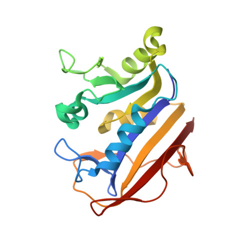 | |
UniProt & NIH Common Fund Data Resources | |||||
PHAROS: P00374 GTEx: ENSG00000228716 | |||||
Entity Groups | |||||
| Sequence Clusters | 30% Identity50% Identity70% Identity90% Identity95% Identity100% Identity | ||||
| UniProt Group | P00374 | ||||
Sequence AnnotationsExpand | |||||
Reference Sequence | |||||
| Ligands 3 Unique | |||||
|---|---|---|---|---|---|
| ID | Chains | Name / Formula / InChI Key | 2D Diagram | 3D Interactions | |
| NAP Download:Ideal Coordinates CCD File | C [auth A], F [auth B] | NADP NICOTINAMIDE-ADENINE-DINUCLEOTIDE PHOSPHATE C21 H28 N7 O17 P3 XJLXINKUBYWONI-NNYOXOHSSA-N |  | ||
| G3V Download:Ideal Coordinates CCD File | D [auth A], G [auth B] | N-(4-{[(2,4-diaminopteridin-6-yl)methyl](hydroxymethyl)amino}benzene-1-carbonyl)-L-glutamic acid C20 H22 N8 O6 KWWKVKGHDTULAH-ZDUSSCGKSA-N |  | ||
| EDO Download:Ideal Coordinates CCD File | E [auth A], H [auth B], I [auth B] | 1,2-ETHANEDIOL C2 H6 O2 LYCAIKOWRPUZTN-UHFFFAOYSA-N |  | ||
| Length ( Å ) | Angle ( ˚ ) |
|---|---|
| a = 37.09 | α = 91.05 |
| b = 42.74 | β = 101.88 |
| c = 72.29 | γ = 120 |
| Software Name | Purpose |
|---|---|
| XDS | data reduction |
| XSCALE | data scaling |
| PHENIX | refinement |
| PDB_EXTRACT | data extraction |
| PHENIX | phasing |
| Coot | model building |
| Funding Organization | Location | Grant Number |
|---|---|---|
| Bill & Melinda Gates Foundation | United States | -- |


