6DCL

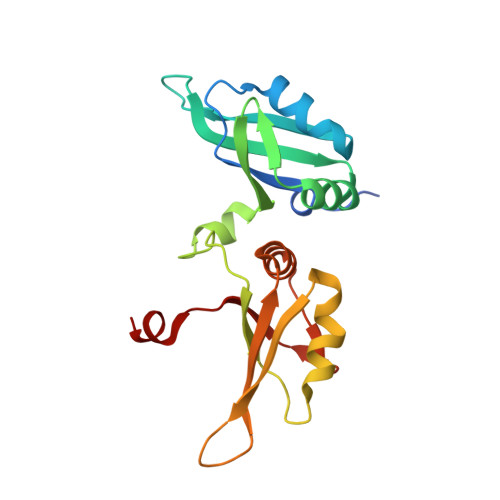
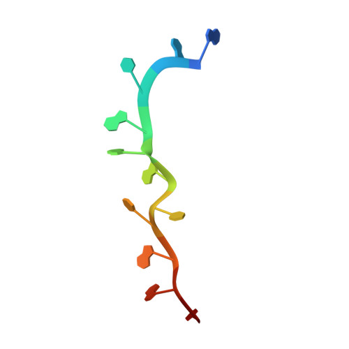
Crystal structure of UP1 bound to pri-miRNA-18a terminal loop


Experimental Data Snapshot

  • Method: X-RAY DIFFRACTION
  • Resolution: 2.50 Å
  • R-Value Free: 0.228 
  • R-Value Work: 0.202 
  • R-Value Observed: 0.203 

wwPDB Validation   3D Report Full Report


This is version 1.3 of the entry. See complete history


Literature

Structural basis for terminal loop recognition and stimulation of pri-miRNA-18a processing by hnRNP A1.

Kooshapur, H.Choudhury, N.R.Simon, B.Muhlbauer, M.Jussupow, A.Fernandez, N.Jones, A.N.Dallmann, A.Gabel, F.Camilloni, C.Michlewski, G.Caceres, J.F.Sattler, M.

(2018) Nat Commun 9: 2479-2479

  • DOI: https://doi.org/10.1038/s41467-018-04871-9
  • Primary Citation of Related Structures:  
    6DCL

  • PubMed Abstract: 

    Post-transcriptional mechanisms play a predominant role in the control of microRNA (miRNA) production. Recognition of the terminal loop of precursor miRNAs by RNA-binding proteins (RBPs) influences their processing; however, the mechanistic basis for how levels of individual or subsets of miRNAs are regulated is mostly unexplored. We previously showed that hnRNP A1, an RBP implicated in many aspects of RNA processing, acts as an auxiliary factor that promotes the Microprocessor-mediated processing of pri-mir-18a. Here, by using an integrative structural biology approach, we show that hnRNP A1 forms a 1:1 complex with pri-mir-18a where both RNA recognition motifs (RRMs) bind to cognate RNA sequence motifs in the terminal loop of pri-mir-18a. Terminal loop binding induces an allosteric destabilization of base-pairing in the pri-mir-18a stem that promotes its downstream processing. Our results highlight terminal loop RNA recognition by RBPs as a potential general principle of miRNA biogenesis and regulation.


  • Organizational Affiliation

    Institute of Structural Biology, Helmholtz Zentrum München, Neuherberg, 85764, Germany.


Macromolecules

Find similar proteins by:  (by identity cutoff)  |  3D Structure
Entity ID: 1
MoleculeChains Sequence LengthOrganismDetailsImage
Heterogeneous nuclear ribonucleoprotein A1
A, B
191Homo sapiensMutation(s): 0 
Gene Names: HNRNPA1HNRPA1
UniProt & NIH Common Fund Data Resources
Find proteins for P09651 (Homo sapiens)
Explore P09651 
Go to UniProtKB:  P09651
PHAROS:  P09651
GTEx:  ENSG00000135486 
Entity Groups  
Sequence Clusters30% Identity50% Identity70% Identity90% Identity95% Identity100% Identity
UniProt GroupP09651
Sequence Annotations
Expand
  • Reference Sequence

Find similar nucleic acids by:  Sequence   |   3D Structure  

Entity ID: 2
MoleculeChains LengthOrganismImage
RNA (5'-R(*AP*GP*UP*AP*GP*AP*UP*UP*AP*GP*C)-3')
C, D
12Homo sapiens
Sequence Annotations
Expand
  • Reference Sequence
Small Molecules
Ligands 1 Unique
IDChains Name / Formula / InChI Key2D Diagram3D Interactions
EDO
Query on EDO

Download Ideal Coordinates CCD File 
E [auth A]1,2-ETHANEDIOL
C2 H6 O2
LYCAIKOWRPUZTN-UHFFFAOYSA-N
Experimental Data & Validation

Experimental Data

  • Method: X-RAY DIFFRACTION
  • Resolution: 2.50 Å
  • R-Value Free: 0.228 
  • R-Value Work: 0.202 
  • R-Value Observed: 0.203 
  • Space Group: P 62 2 2
Unit Cell:
Length ( Å )Angle ( ˚ )
a = 127.028α = 90
b = 127.028β = 90
c = 147.064γ = 120
Software Package:
Software NamePurpose
PHENIXrefinement
Aimlessdata scaling
PDB_EXTRACTdata extraction
XDSdata reduction
PHASERphasing

Structure Validation

View Full Validation Report


Entry History & Funding Information

Deposition Data


Funding OrganizationLocationGrant Number
German Research Foundation (DFG)GermanySFB1035
German Research Foundation (DFG)GermanyGRK1721

Revision History  (Full details and data files)

  • Version 1.0: 2018-06-27
    Type: Initial release
  • Version 1.1: 2018-07-11
    Changes: Data collection, Database references
  • Version 1.2: 2020-01-08
    Changes: Author supporting evidence
  • Version 1.3: 2024-03-13
    Changes: Data collection, Database references, Refinement description