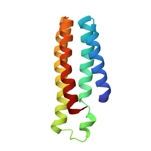
6DYG

Fe(II)-bound structure of the engineered cyt cb562 variant, CH3Y


Experimental Data Snapshot

  • Method: X-RAY DIFFRACTION
  • Resolution: 1.49 Å
  • R-Value Free: 0.217 
  • R-Value Work: 0.191 
  • R-Value Observed: 0.192 

Starting Model: experimental
View more details

wwPDB Validation   3D Report Full Report


This is version 1.5 of the entry. See complete history


Literature

An efficient, step-economical strategy for the design of functional metalloproteins.

Rittle, J.Field, M.J.Green, M.T.Tezcan, F.A.

(2019) Nat Chem 11: 434-441

  • DOI: https://doi.org/10.1038/s41557-019-0218-9
  • Primary Citation of Related Structures:  
    6DY4, 6DY6, 6DY8, 6DYB, 6DYC, 6DYD, 6DYE, 6DYF, 6DYG, 6DYH, 6DYI, 6DYJ, 6DYK, 6DYL

  • PubMed Abstract: 

    The bottom-up design and construction of functional metalloproteins remains a formidable task in biomolecular design. Although numerous strategies have been used to create new metalloproteins, pre-existing knowledge of the tertiary and quaternary protein structure is often required to generate suitable platforms for robust metal coordination and activity. Here we report an alternative and easily implemented approach (metal active sites by covalent tethering or MASCoT) in which folded protein building blocks are linked by a single disulfide bond to create diverse metal coordination environments within evolutionarily naive protein-protein interfaces. Metalloproteins generated using this strategy uniformly bind a wide array of first-row transition metal ions (Mn II , Fe II , Co II , Ni II , Cu II , Zn II and vanadyl) with physiologically relevant thermodynamic affinities (dissociation constants ranging from 700 nM for Mn II to 50 fM for Cu II ). MASCoT readily affords coordinatively unsaturated metal centres-including a penta-His-coordinated non-haem Fe site-and well-defined binding pockets that can accommodate modifications and enable coordination of exogenous ligands such as nitric oxide to the interfacial metal centre.


  • Organizational Affiliation

    Department of Chemistry and Biochemistry, University of California, San Diego, La Jolla, CA, USA.


Macromolecules
Find similar proteins by:  (by identity cutoff)  |  3D Structure
Entity ID: 1
MoleculeChains Sequence LengthOrganismDetailsImage
Soluble cytochrome b562
A, B
106Escherichia coliMutation(s): 8 
Gene Names: cybC
UniProt
Find proteins for P0ABE7 (Escherichia coli)
Explore P0ABE7 
Go to UniProtKB:  P0ABE7
Entity Groups  
Sequence Clusters30% Identity50% Identity70% Identity90% Identity95% Identity100% Identity
UniProt GroupP0ABE7
Sequence Annotations
Expand
  • Reference Sequence
Experimental Data & Validation

Experimental Data

  • Method: X-RAY DIFFRACTION
  • Resolution: 1.49 Å
  • R-Value Free: 0.217 
  • R-Value Work: 0.191 
  • R-Value Observed: 0.192 
  • Space Group: P 1 21 1
Unit Cell:
Length ( Å )Angle ( ˚ )
a = 34.739α = 90
b = 83.513β = 99.99
c = 39.088γ = 90
Software Package:
Software NamePurpose
PHENIXrefinement
MOSFLMdata reduction
Aimlessdata scaling
PHASERphasing

Structure Validation

View Full Validation Report


Entry History & Funding Information

Deposition Data


Funding OrganizationLocationGrant Number
National Institutes of Health/National Institute of General Medical Sciences (NIH/NIGMS)United StatesGM120981
National Science Foundation (NSF, United States)United StatesCHE1607145

Revision History  (Full details and data files)

  • Version 1.0: 2019-04-24
    Type: Initial release
  • Version 1.1: 2019-05-08
    Changes: Data collection, Database references
  • Version 1.2: 2019-11-20
    Changes: Database references
  • Version 1.3: 2019-11-27
    Changes: Author supporting evidence
  • Version 1.4: 2023-10-11
    Changes: Data collection, Database references, Derived calculations, Refinement description
  • Version 1.5: 2024-10-16
    Changes: Structure summary