Origins of glycan selectivity in streptococcal Siglec-like adhesins suggest mechanisms of receptor adaptation.
Bensing, B.A., Stubbs, H.E., Agarwal, R., Yamakawa, I., Luong, K., Solakyildirim, K., Yu, H., Hadadianpour, A., Castro, M.A., Fialkowski, K.P., Morrison, K.M., Wawrzak, Z., Chen, X., Lebrilla, C.B., Baudry, J., Smith, J.C., Sullam, P.M., Iverson, T.M.(2022) Nat Commun 13: 2753-2753
- PubMed: 35585145 Search on PubMedSearch on PubMed Central
- DOI: https://doi.org/10.1038/s41467-022-30509-y
- Primary Citation Related Structures:
6EF7, 6EF9, 6EFA, 6EFB, 6EFC, 6EFD, 6EFF, 6EFI, 6X3K, 6X3Q, 7KMJ - PubMed Abstract:
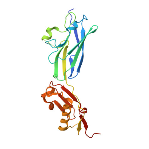
Bacterial binding to host receptors underlies both commensalism and pathogenesis. Many streptococci adhere to protein-attached carbohydrates expressed on cell surfaces using Siglec-like binding regions (SLBRs). The precise glycan repertoire recognized may dictate whether the organism is a strict commensal versus a pathogen. However, it is currently not clear what drives receptor selectivity. Here, we use five representative SLBRs and identify regions of the receptor binding site that are hypervariable in sequence and structure. We show that these regions control the identity of the preferred carbohydrate ligand using chimeragenesis and single amino acid substitutions. We further evaluate how the identity of the preferred ligand affects the interaction with glycoprotein receptors in human saliva and plasma samples. As point mutations can change the preferred human receptor, these studies suggest how streptococci may adapt to changes in the environmental glycan repertoire.
- Division of Infectious Diseases, Veterans Affairs Medical Center, Department of Medicine, University of California, San Francisco, CA, USA.
Organizational Affiliation: