6EIC | pdb_00006eic

Crystal structure of Rv0183, a Monoglyceride Lipase from Mycobacterium Tuberculosis


Experimental Data Snapshot

  • Method: X-RAY DIFFRACTION
  • Resolution: 1.80 Å
  • R-Value Free: 
    0.251 (Depositor), 0.271 (DCC) 
  • R-Value Work: 
    0.221 (Depositor), 0.241 (DCC) 
  • R-Value Observed: 
    0.223 (Depositor) 

wwPDB Validation 3D Report Full Report

Validation slider image for 6EIC

This is version 1.2 of the entry. See complete history

Literature

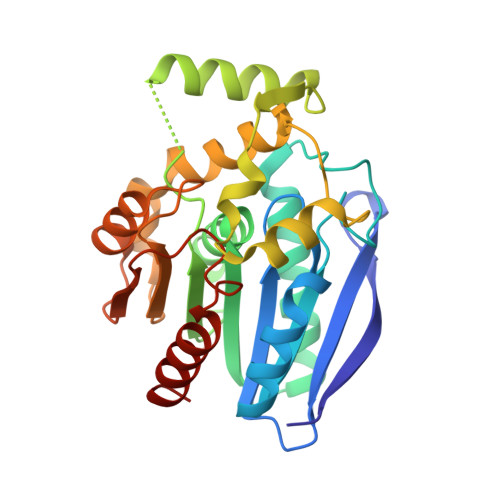
The crystal structure of monoacylglycerol lipase from M. tuberculosis reveals the basis for specific inhibition.

Aschauer, P.Zimmermann, R.Breinbauer, R.Pavkov-Keller, T.Oberer, M.

(2018) Sci Rep 8: 8948-8948

  • DOI: https://doi.org/10.1038/s41598-018-27051-7
  • Primary Citation Related Structures: 
    6EIC

  • PubMed Abstract: 

    Monoacylglycerol lipases (MGLs) are enzymes that hydrolyze monoacylglycerol into a free fatty acid and glycerol. Fatty acids can be used for triacylglycerol synthesis, as energy source, as building blocks for energy storage, and as precursor for membrane phospholipids. In Mycobacterium tuberculosis, fatty acids also serve as precursor for polyketide lipids like mycolic acids, major components of the cellular envelope associated to resistance for drug. We present the crystal structure of the MGL Rv0183 from Mycobacterium tuberculosis (mtbMGL) in open conformation. The structure reveals remarkable similarities with MGL from humans (hMGL) in both, the cap region and the α/β core. Nevertheless, mtbMGL could not be inhibited with JZL-184, a known inhibitor of hMGL. Docking studies provide an explanation why the activity of mtbMGL was not affected by the inhibitor. Our findings suggest that specific inhibition of mtbMGL from Mycobacterium tuberculosis, one of the oldest recognized pathogens, is possible without influencing hMGL.


  • Organizational Affiliation
    • Institute of Molecular Biosciences, University of Graz, Humboldtstraße 50/3, 8010, Graz, Austria.

Macromolecule Content 

  • Total Structure Weight: 91.41 kDa 
  • Atom Count: 6,809 
  • Modeled Residue Count: 826 
  • Deposited Residue Count: 837 
  • Unique protein chains: 1

Macromolecules

Find similar proteins by:|  3D Structure
Entity ID: 1
MoleculeChains  Sequence LengthOrganismDetailsImage
Mycobacterium Tuberculosis Monoglyceride LipaseA [auth C],
B [auth A],
C [auth B]
279Mycobacterium tuberculosis H37RvMutation(s): 0 
Gene Names: Rv0183
EC: 3.1.1.23
UniProt
Find proteins for O07427 (Mycobacterium tuberculosis (strain ATCC 25618 / H37Rv))
Explore O07427 
Go to UniProtKB:  O07427
Entity Groups
Sequence Clusters30% Identity50% Identity70% Identity90% Identity95% Identity100% Identity
UniProt GroupO07427
Sequence Annotations
Expand
Reference Sequence

Experimental Data & Validation

Experimental Data

  • Method: X-RAY DIFFRACTION
  • Resolution: 1.80 Å
  • R-Value Free:  0.251 (Depositor), 0.271 (DCC) 
  • R-Value Work:  0.221 (Depositor), 0.241 (DCC) 
  • R-Value Observed: 0.223 (Depositor) 
Space Group: P 31 1 2
Unit Cell:
Length ( Å )Angle ( ˚ )
a = 85.825α = 90
b = 85.825β = 90
c = 196.98γ = 120
Software Package:
Software NamePurpose
PHENIXrefinement
SCALAdata reduction
SCALAdata scaling
BALBESphasing

Structure Validation

View Full Validation Report


Entry History 

& Funding Information

Deposition Data


Funding OrganizationLocationGrant Number
Austrian Science FundAustriaP 24857

Revision History  (Full details and data files)

  • Version 1.0: 2018-06-27
    Type: Initial release
  • Version 1.1: 2021-09-29
    Changes: Database references, Structure summary
  • Version 1.2: 2024-06-19
    Changes: Data collection