6EIJ | pdb_00006eij

DYRK1A in complex with HG-8-60-1


Experimental Data Snapshot

  • Method: X-RAY DIFFRACTION
  • Resolution: 2.42 Å
  • R-Value Free: 
    0.242 (Depositor), 0.244 (DCC) 
  • R-Value Work: 
    0.213 (Depositor), 0.214 (DCC) 
  • R-Value Observed: 
    0.215 (Depositor) 

Starting Model: experimental
View more details

wwPDB Validation 3D Report Full Report

Validation slider image for 6EIJ

Ligand Structure Quality Assessment 


This is version 1.3 of the entry. See complete history

Literature

Novel Scaffolds for Dual Specificity Tyrosine-Phosphorylation-Regulated Kinase (DYRK1A) Inhibitors.

Czarna, A.Wang, J.Zelencova, D.Liu, Y.Deng, X.Choi, H.G.Zhang, T.Zhou, W.Chang, J.W.Kildalsen, H.Seternes, O.M.Gray, N.S.Engh, R.A.Rothweiler, U.

(2018) J Med Chem 61: 7560-7572

  • DOI: https://doi.org/10.1021/acs.jmedchem.7b01847
  • Primary Citation Related Structures: 
    6EIF, 6EIJ, 6EIL, 6EIP, 6EIQ, 6EIR, 6EIS, 6EIV, 6EJ4

  • PubMed Abstract: 

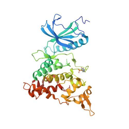
    DYRK1A is one of five members of the dual-specificity tyrosine (Y) phosphorylation-regulated kinase (DYRK) family. The DYRK1A gene is located in the Down syndrome critical region and regulates cellular processes related to proliferation and differentiation of neuronal progenitor cells during early development. This has focused research on its role in neuronal degenerative diseases, including Alzheimer's and Down syndrome. Recent studies have also shown a possible role of DYRK1A in diabetes. Here we report a variety of scaffolds not generally known for DYRK1A inhibition, demonstrating their effects in in vitro assays and also in cell cultures. These inhibitors effectively block the tau phosphorylation that is a hallmark of Alzheimer's disease. The crystal structures of these inhibitors support the design of optimized and novel therapeutics.


  • Organizational Affiliation
    • Department of Pharmacy, Faculty of Health Sciences , UiT The Arctic University of Norway , N-9037 Tromsø , Norway.

Macromolecule Content 

  • Total Structure Weight: 172.48 kDa 
  • Atom Count: 11,489 
  • Modeled Residue Count: 1,359 
  • Deposited Residue Count: 1,472 
  • Unique protein chains: 1

Macromolecules

Find similar proteins by:|  3D Structure
Entity ID: 1
MoleculeChains  Sequence LengthOrganismDetailsImage
Dual specificity tyrosine-phosphorylation-regulated kinase 1A
A, B, C, D
368Homo sapiensMutation(s): 0 
Gene Names: DYRK1ADYRKMNBMNBH
EC: 2.7.12.1 (PDB Primary Data), 2.7.11.23 (UniProt)
UniProt & NIH Common Fund Data Resources
Find proteins for Q13627 (Homo sapiens)
Explore Q13627 
Go to UniProtKB:  Q13627
PHAROS:  Q13627
GTEx:  ENSG00000157540 
Entity Groups
Sequence Clusters30% Identity50% Identity70% Identity90% Identity95% Identity100% Identity
UniProt GroupQ13627
Sequence Annotations
Expand
Reference Sequence

Small Molecules

Experimental Data & Validation

Experimental Data

  • Method: X-RAY DIFFRACTION
  • Resolution: 2.42 Å
  • R-Value Free:  0.242 (Depositor), 0.244 (DCC) 
  • R-Value Work:  0.213 (Depositor), 0.214 (DCC) 
  • R-Value Observed: 0.215 (Depositor) 
Space Group: P 21 21 21
Unit Cell:
Length ( Å )Angle ( ˚ )
a = 89.24α = 90
b = 88.04β = 90
c = 228.5γ = 90
Software Package:
Software NamePurpose
REFMACrefinement
XDSdata reduction
XDSdata scaling
PHASERphasing

Structure Validation

View Full Validation Report


Ligand Structure Quality Assessment 


Entry History 

Deposition Data

Revision History  (Full details and data files)

  • Version 1.0: 2018-08-29
    Type: Initial release
  • Version 1.1: 2018-09-26
    Changes: Data collection, Database references
  • Version 1.2: 2024-01-17
    Changes: Data collection, Database references, Refinement description
  • Version 1.3: 2024-10-16
    Changes: Structure summary