6EWA | pdb_00006ewa

Crystal structure of HLA-A2 in complex with LILRB1


Experimental Data Snapshot

  • Method: X-RAY DIFFRACTION
  • Resolution: 2.39 Å
  • R-Value Free: 
    0.279 (Depositor), 0.276 (DCC) 
  • R-Value Work: 
    0.228 (Depositor), 0.232 (DCC) 
  • R-Value Observed: 
    0.231 (Depositor) 

Starting Model: experimental
View more details

wwPDB Validation 3D Report Full Report

Validation slider image for 6EWA

This is version 1.3 of the entry. See complete history

Literature

Application of the immunoregulatory receptor LILRB1 as a crystallisation chaperone for human class I MHC complexes.

Mohammed, F.Stones, D.H.Willcox, B.E.

(2019) J Immunol Methods 464: 47-56

  • DOI: https://doi.org/10.1016/j.jim.2018.10.011
  • Primary Citation Related Structures: 
    6EWA, 6EWC, 6EWO

  • PubMed Abstract: 

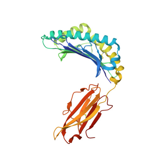
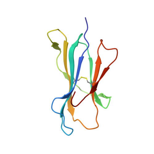
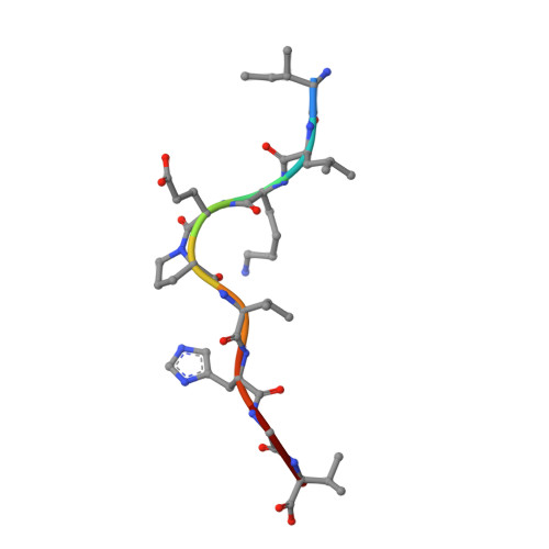
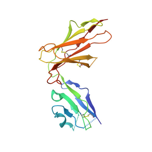
    X-ray crystallographic studies of class I peptide-MHC molecules (pMHC) continue to provide important insights into immune recognition, however their success depends on generation of diffraction-quality crystals, which remains a significant challenge. While protein engineering techniques such as surface-entropy reduction and lysine methylation have proven utility in facilitating and/or improving protein crystallisation, they risk affecting the conformation and biochemistry of the class I MHC antigen binding groove. An attractive alternative is the use of noncovalent crystallisation chaperones, however these have not been developed for pMHC. Here we describe a method for promoting class I pMHC crystallisation, by exploiting its natural ligand interaction with the immunoregulatory receptor LILRB1 as a novel crystallisation chaperone. First, focussing on a model HIV-1-derived HLA-A2-restricted peptide, we determined a 2.4 Å HLA-A2/LILRB1 structure, which validated that co-crystallisation with LILRB1 does not alter conformation of the antigenic peptide. We then demonstrated that addition of LILRB1 enhanced the crystallisation of multiple peptide-HLA-A2 complexes, and identified a generic condition for initial co-crystallisation. LILRB1 chaperone-based crystallisation enabled structure determination for HLA-A2 complexes previously intransigent to crystallisation, including both conventional and post-translationally-modified peptides, of diverse lengths. Since both the LILRB1 recognition interface on the HLA-A2 α3 domain molecule and HLA-A2-mediated crystal contacts are predominantly conserved across class I MHC molecules, the approach we outline could prove applicable to a diverse range of class I pMHC. LILRB1 chaperone-mediated crystallisation should expedite molecular insights into the immunobiology of diverse immune-related diseases and immunotherapeutic strategies, particularly involving class I pMHC complexes that are challenging to crystallise.


  • Organizational Affiliation
    • Cancer Immunology and Immunotherapy Centre, Institute of Immunology and Immunotherapy, University of Birmingham, Edgbaston, Birmingham B15 2TT, UK.

Macromolecule Content 

  • Total Structure Weight: 132.88 kDa 
  • Atom Count: 9,148 
  • Modeled Residue Count: 1,123 
  • Deposited Residue Count: 1,158 
  • Unique protein chains: 4

Macromolecules

Find similar proteins by:|  3D Structure
Entity ID: 1
MoleculeChains  Sequence LengthOrganismDetailsImage
HLA class I histocompatibility antigen, A-2 alpha chain
A, E
276Homo sapiensMutation(s): 0 
Gene Names: HLA-AHLAA
UniProt & NIH Common Fund Data Resources
Find proteins for P04439 (Homo sapiens)
Explore P04439 
Go to UniProtKB:  P04439
PHAROS:  P04439
GTEx:  ENSG00000206503 
Entity Groups
Sequence Clusters30% Identity50% Identity70% Identity90% Identity95% Identity100% Identity
UniProt GroupP04439
Sequence Annotations
Expand
Reference Sequence
Find similar proteins by:|  3D Structure
Entity ID: 2
MoleculeChains  Sequence LengthOrganismDetailsImage
Beta-2-microglobulin
B, F
99Homo sapiensMutation(s): 0 
Gene Names: B2MCDABP0092HDCMA22P
UniProt & NIH Common Fund Data Resources
Find proteins for P61769 (Homo sapiens)
Explore P61769 
Go to UniProtKB:  P61769
PHAROS:  P61769
GTEx:  ENSG00000166710 
Entity Groups
Sequence Clusters30% Identity50% Identity70% Identity90% Identity95% Identity100% Identity
UniProt GroupP61769
Sequence Annotations
Expand
Reference Sequence
Find similar proteins by:  Sequence   |   3D Structure  
Entity ID: 3
MoleculeChains  Sequence LengthOrganismDetailsImage
Polyprotein
C, G
9Human immunodeficiency virus 1Mutation(s): 0 
UniProt
Find proteins for P04585 (Human immunodeficiency virus type 1 group M subtype B (isolate HXB2))
Explore P04585 
Go to UniProtKB:  P04585
Entity Groups
UniProt GroupP04585
Sequence Annotations
Expand
Reference Sequence
Find similar proteins by:|  3D Structure
Entity ID: 4
MoleculeChains  Sequence LengthOrganismDetailsImage
LIR-1
D, H
195Homo sapiensMutation(s): 0 
Gene Names: LILRB1
UniProt & NIH Common Fund Data Resources
Find proteins for Q8NHL6 (Homo sapiens)
Explore Q8NHL6 
Go to UniProtKB:  Q8NHL6
PHAROS:  Q8NHL6
Entity Groups
Sequence Clusters30% Identity50% Identity70% Identity90% Identity95% Identity100% Identity
UniProt GroupQ8NHL6
Sequence Annotations
Expand
Reference Sequence

Experimental Data & Validation

Experimental Data

  • Method: X-RAY DIFFRACTION
  • Resolution: 2.39 Å
  • R-Value Free:  0.279 (Depositor), 0.276 (DCC) 
  • R-Value Work:  0.228 (Depositor), 0.232 (DCC) 
  • R-Value Observed: 0.231 (Depositor) 
Space Group: P 32 2 1
Unit Cell:
Length ( Å )Angle ( ˚ )
a = 116.15α = 90
b = 116.15β = 90
c = 192.82γ = 120
Software Package:
Software NamePurpose
SCALAdata scaling
REFMACrefinement
PDB_EXTRACTdata extraction
MOSFLMdata reduction
MOLREPphasing

Structure Validation

View Full Validation Report


Entry History 

Deposition Data

Revision History  (Full details and data files)

  • Version 1.0: 2018-11-07
    Type: Initial release
  • Version 1.1: 2019-01-16
    Changes: Data collection, Database references
  • Version 1.2: 2024-01-17
    Changes: Advisory, Data collection, Database references, Refinement description
  • Version 1.3: 2024-11-13
    Changes: Structure summary