6FJJ | pdb_00006fjj

Joint neutron and x-ray crystal structure of human carbonic anhydrase IX mimic (saccharin).


Experimental Data Snapshot

  • Method: X-RAY DIFFRACTION
  • Resolution: 1.50 Å
  • R-Value Free: 
    0.166 (Depositor), 0.174 (DCC) 
  • R-Value Work: 
    0.135 (Depositor), 0.165 (DCC) 
  • R-Value Observed: 
    0.137 (Depositor) 

  • Method: NEUTRON DIFFRACTION
  • Resolution: 2.00 Å
  • R-Value Free: 
    0.231 (Depositor) 
  • R-Value Work: 
    0.205 (Depositor) 
  • R-Value Observed: 
    0.207 (Depositor) 

Starting Model: experimental
View more details

wwPDB Validation 3D Report Full Report

Validation slider image for 6FJJ

This is version 1.2 of the entry. See complete history

Literature

Using neutron crystallography to elucidate the basis of selective inhibition of carbonic anhydrase by saccharin and a derivative.

Koruza, K.Mahon, B.P.Blakeley, M.P.Ostermann, A.Schrader, T.E.McKenna, R.Knecht, W.Fisher, S.Z.

(2019) J Struct Biol 205: 147-154

  • DOI: https://doi.org/10.1016/j.jsb.2018.12.009
  • Primary Citation Related Structures: 
    6FJI, 6FJJ, 6GCY

  • PubMed Abstract: 

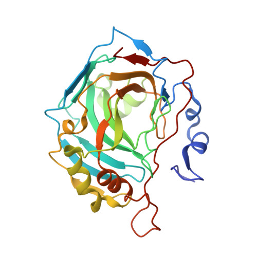
    Up-regulation of carbonic anhydrase IX (CA IX) expression is an indicator of metastasis and associated with poor cancer patient prognosis. CA IX has emerged as a cancer drug target but development of isoform-specific inhibitors is challenging due to other highly conserved CA isoforms. In this study, a CA IX mimic construct was used (CA II with seven point mutations introduced, to mimic CA IX active site) while maintaining CA II solubility that make it amenable to crystallography. The structures of CA IX mimic unbound and in complex with saccharin (SAC) and a saccharin-glucose conjugate (SGC) were determined using joint X-ray and neutron protein crystallography. Previously, SAC and SGC have been shown to display CA isoform inhibitor selectivity in assays and X-ray crystal structures failed to reveal the basis of this selectivity. Joint X-ray and neutron crystallographic studies have shown active site residues, solvent, and H-bonding re-organization upon SAC and SGC binding. These observations highlighted the importance of residues 67 (Asn in CA II, Gln in CA IX) and 130 (Asp in CA II, Arg in CA IX) in selective CA inhibitor targeting.


  • Organizational Affiliation
    • Lund Protein Production Platform (LP3) & Department of Biology, Lund University, 223 62 Lund, Sweden.

Macromolecule Content 

  • Total Structure Weight: 29.45 kDa 
  • Atom Count: 2,287 
  • Modeled Residue Count: 258 
  • Deposited Residue Count: 260 
  • Unique protein chains: 1

Macromolecules

Find similar proteins by:|  3D Structure
Entity ID: 1
MoleculeChains  Sequence LengthOrganismDetailsImage
Carbonic anhydrase 2260Homo sapiensMutation(s): 7 
Gene Names: CA2
EC: 4.2.1.1 (PDB Primary Data), 4.2.1.69 (UniProt)
UniProt & NIH Common Fund Data Resources
Find proteins for P00918 (Homo sapiens)
Explore P00918 
Go to UniProtKB:  P00918
PHAROS:  P00918
GTEx:  ENSG00000104267 
Entity Groups
Sequence Clusters30% Identity50% Identity70% Identity90% Identity95% Identity100% Identity
UniProt GroupP00918
Sequence Annotations
Expand
Reference Sequence

Small Molecules

Ligands 2 Unique
IDChains Name / Formula / InChI Key2D Diagram3D Interactions
LSA

Query on LSA


Download:Ideal Coordinates CCD File
C [auth A]1,2-BENZISOTHIAZOL-3(2H)-ONE 1,1-DIOXIDE
C7 H5 N O3 S
CVHZOJJKTDOEJC-UHFFFAOYSA-N
ZN

Query on ZN


Download:Ideal Coordinates CCD File
B [auth A]ZINC ION
Zn
PTFCDOFLOPIGGS-UHFFFAOYSA-N

Experimental Data & Validation

Experimental Data

  • Method: X-RAY DIFFRACTION
  • Resolution: 1.50 Å
  • R-Value Free:  0.166 (Depositor), 0.174 (DCC) 
  • R-Value Work:  0.135 (Depositor), 0.165 (DCC) 
  • R-Value Observed: 0.137 (Depositor) 
  • Method: NEUTRON DIFFRACTION
  • Resolution: 2.00 Å
  • R-Value Free:  0.231 (Depositor) 
  • R-Value Work:  0.205 (Depositor) 
  • R-Value Observed: 0.207 (Depositor) 
Space Group: P 1 21 1
Unit Cell:
Length ( Å )Angle ( ˚ )
a = 42.53α = 90
b = 41.83β = 104.04
c = 72.7γ = 90
Software Package:
Software NamePurpose
PHENIXrefinement
XDSdata reduction
XDSdata scaling

Structure Validation

View Full Validation Report


Entry History 

Deposition Data

  • Released Date: 2019-02-06 
  • Deposition Author(s): Fisher, S.Z.

Revision History  (Full details and data files)

  • Version 1.0: 2019-02-06
    Type: Initial release
  • Version 1.1: 2019-03-06
    Changes: Data collection, Database references
  • Version 1.2: 2024-05-01
    Changes: Data collection, Database references, Refinement description