6FU5 | pdb_00006fu5

Structure of the kinase domain of human RIPK2 in complex with the inhibitor CSLP18


Experimental Data Snapshot

  • Method: X-RAY DIFFRACTION
  • Resolution: 3.26 Å
  • R-Value Free: 
    0.271 (Depositor), 0.277 (DCC) 
  • R-Value Work: 
    0.229 (Depositor), 0.231 (DCC) 
  • R-Value Observed: 
    0.231 (Depositor) 

Starting Model: experimental
View more details

wwPDB Validation 3D Report Full Report

Validation slider image for 6FU5

Ligand Structure Quality Assessment 


This is version 1.4 of the entry. See complete history

Literature

Small molecule inhibitors reveal an indispensable scaffolding role of RIPK2 in NOD2 signaling.

Hrdinka, M.Schlicher, L.Dai, B.Pinkas, D.M.Bufton, J.C.Picaud, S.Ward, J.A.Rogers, C.Suebsuwong, C.Nikhar, S.Cuny, G.D.Huber, K.V.Filippakopoulos, P.Bullock, A.N.Degterev, A.Gyrd-Hansen, M.

(2018) EMBO J 37

  • DOI: https://doi.org/10.15252/embj.201899372
  • Primary Citation Related Structures: 
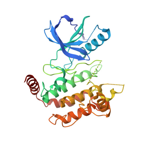
    6FU5

  • PubMed Abstract: 

    RIPK2 mediates inflammatory signaling by the bacteria-sensing receptors NOD1 and NOD2. Kinase inhibitors targeting RIPK2 are a proposed strategy to ameliorate NOD-mediated pathologies. Here, we reveal that RIPK2 kinase activity is dispensable for NOD2 inflammatory signaling and show that RIPK2 inhibitors function instead by antagonizing XIAP-binding and XIAP-mediated ubiquitination of RIPK2. We map the XIAP binding site on RIPK2 to the loop between β2 and β3 of the N-lobe of the kinase, which is in close proximity to the ATP-binding pocket. Through characterization of a new series of ATP pocket-binding RIPK2 inhibitors, we identify the molecular features that determine their inhibition of both the RIPK2-XIAP interaction, and of cellular and in vivo NOD2 signaling. Our study exemplifies how targeting of the ATP-binding pocket in RIPK2 can be exploited to interfere with the RIPK2-XIAP interaction for modulation of NOD signaling.


  • Organizational Affiliation
    • Nuffield Department of Clinical Medicine, Ludwig Institute for Cancer Research, University of Oxford, Oxford, UK.

Macromolecule Content 

  • Total Structure Weight: 73.73 kDa 
  • Atom Count: 4,634 
  • Modeled Residue Count: 568 
  • Deposited Residue Count: 634 
  • Unique protein chains: 1

Macromolecules

Find similar proteins by:|  3D Structure
Entity ID: 1
MoleculeChains  Sequence LengthOrganismDetailsImage
Receptor-interacting serine/threonine-protein kinase 2
A, B
317Homo sapiensMutation(s): 0 
Gene Names: RIPK2CARDIAKRICKRIP2UNQ277/PRO314/PRO34092
EC: 2.7.11.1 (PDB Primary Data), 2.7.10.2 (PDB Primary Data)
UniProt & NIH Common Fund Data Resources
Find proteins for O43353 (Homo sapiens)
Explore O43353 
Go to UniProtKB:  O43353
PHAROS:  O43353
GTEx:  ENSG00000104312 
Entity Groups
Sequence Clusters30% Identity50% Identity70% Identity90% Identity95% Identity100% Identity
UniProt GroupO43353
Sequence Annotations
Expand
Reference Sequence

Small Molecules

Ligands 1 Unique
IDChains Name / Formula / InChI Key2D Diagram3D Interactions
E7N
(Subject of Investigation/LOI)

Query on E7N


Download:Ideal Coordinates CCD File
C [auth A],
D [auth B]
~{N}-[5-[2-azanyl-5-(4-piperazin-1-ylphenyl)pyridin-3-yl]-2-methoxy-phenyl]propane-1-sulfonamide
C25 H31 N5 O3 S
JMJHOMFUECZGAD-UHFFFAOYSA-N

Experimental Data & Validation

Experimental Data

  • Method: X-RAY DIFFRACTION
  • Resolution: 3.26 Å
  • R-Value Free:  0.271 (Depositor), 0.277 (DCC) 
  • R-Value Work:  0.229 (Depositor), 0.231 (DCC) 
  • R-Value Observed: 0.231 (Depositor) 
Space Group: P 21 21 21
Unit Cell:
Length ( Å )Angle ( ˚ )
a = 75.79α = 90
b = 122.71β = 90
c = 71.45γ = 90
Software Package:
Software NamePurpose
PHENIXrefinement
xia2data reduction
xia2data scaling
PHASERphasing

Structure Validation

View Full Validation Report


Ligand Structure Quality Assessment 


Entry History 

Deposition Data

Revision History  (Full details and data files)

  • Version 1.0: 2018-04-25
    Type: Initial release
  • Version 1.1: 2018-08-01
    Changes: Data collection, Database references
  • Version 1.2: 2018-08-29
    Changes: Data collection, Structure summary
  • Version 1.3: 2018-09-12
    Changes: Data collection, Database references
  • Version 1.4: 2024-01-17
    Changes: Data collection, Database references, Refinement description