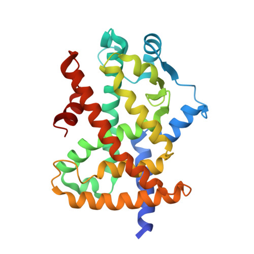
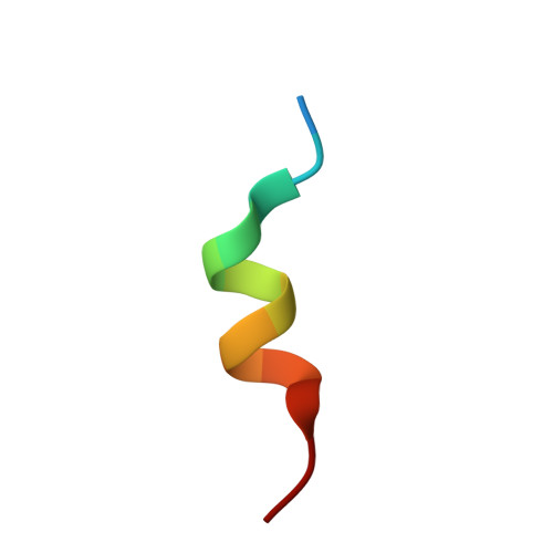
6FZF

PPAR mutant complex


Experimental Data Snapshot

  • Method: X-RAY DIFFRACTION
  • Resolution: 1.95 Å
  • R-Value Free: 0.194 
  • R-Value Work: 0.152 
  • R-Value Observed: 0.153 

Starting Model: experimental
View more details

wwPDB Validation   3D Report Full Report


Ligand Structure Quality Assessment 


This is version 1.1 of the entry. See complete history


Literature

Recurrent activating mutations of PPAR gamma associated with luminal bladder tumors.

Rochel, N.Krucker, C.Coutos-Thevenot, L.Osz, J.Zhang, R.Guyon, E.Zita, W.Vanthong, S.Hernandez, O.A.Bourguet, M.Badawy, K.A.Dufour, F.Peluso-Iltis, C.Heckler-Beji, S.Dejaegere, A.Kamoun, A.de Reynies, A.Neuzillet, Y.Rebouissou, S.Beraud, C.Lang, H.Massfelder, T.Allory, Y.Cianferani, S.Stote, R.H.Radvanyi, F.Bernard-Pierrot, I.

(2019) Nat Commun 10: 253-253

  • DOI: https://doi.org/10.1038/s41467-018-08157-y
  • Primary Citation of Related Structures:  
    6FZF, 6FZG, 6FZJ, 6FZP, 6FZY

  • PubMed Abstract: 

    The upregulation of PPARγ/RXRα transcriptional activity has emerged as a key event in luminal bladder tumors. It renders tumor cell growth PPARγ-dependent and modulates the tumor microenvironment to favor escape from immuno-surveillance. The activation of the pathway has been linked to PPARG gains/amplifications resulting in PPARγ overexpression and to recurrent activating point mutations of RXRα. Here, we report recurrent mutations of PPARγ that also activate the PPARγ/RXRα pathway, conferring PPARγ-dependency and supporting a crucial role of PPARγ in luminal bladder cancer. These mutations are found throughout the protein-including N-terminal, DNA-binding and ligand-binding domains-and most of them enhance protein activity. Structure-function studies of PPARγ variants with mutations in the ligand-binding domain allow identifying structural elements that underpin their gain-of-function. Our study reveals genomic alterations of PPARG that lead to pro-tumorigenic PPARγ/RXRα pathway activation in luminal bladder tumors and may open the way towards alternative options for treatment.


  • Organizational Affiliation

    Institut de Génétique et de Biologie Moléculaire et Cellulaire (IGBMC), Institut National de La Santé et de La Recherche Médicale (INSERM), U1258/Centre National de Recherche Scientifique (CNRS), UMR7104/Université de Strasbourg, 67404 Illkirch, France. rochel@igbmc.fr.


Macromolecules
Find similar proteins by:  (by identity cutoff)  |  3D Structure
Entity ID: 1
MoleculeChains Sequence LengthOrganismDetailsImage
Peroxisome proliferator-activated receptor gamma
A, B
279Homo sapiensMutation(s): 1 
Gene Names: PPARGNR1C3
UniProt & NIH Common Fund Data Resources
Find proteins for P37231 (Homo sapiens)
Explore P37231 
Go to UniProtKB:  P37231
PHAROS:  P37231
GTEx:  ENSG00000132170 
Entity Groups  
Sequence Clusters30% Identity50% Identity70% Identity90% Identity95% Identity100% Identity
UniProt GroupP37231
Sequence Annotations
Expand
  • Reference Sequence

Find similar proteins by:  Sequence   |   3D Structure  

Entity ID: 2
MoleculeChains Sequence LengthOrganismDetailsImage
Peroxisome proliferator-activated receptor gamma coactivator 1-alpha
C, D
14Homo sapiensMutation(s): 0 
UniProt & NIH Common Fund Data Resources
Find proteins for Q9UBK2 (Homo sapiens)
Explore Q9UBK2 
Go to UniProtKB:  Q9UBK2
PHAROS:  Q9UBK2
GTEx:  ENSG00000109819 
Entity Groups  
Sequence Clusters30% Identity50% Identity70% Identity90% Identity95% Identity100% Identity
UniProt GroupQ9UBK2
Sequence Annotations
Expand
  • Reference Sequence
Small Molecules
Ligands 1 Unique
IDChains Name / Formula / InChI Key2D Diagram3D Interactions
EDK
Query on EDK

Download Ideal Coordinates CCD File 
E [auth A],
F [auth B]
(2~{S})-3-[4-[2-[methyl(pyridin-2-yl)amino]ethoxy]phenyl]-2-[[2-(phenylcarbonyl)phenyl]amino]propanoic acid
C30 H29 N3 O4
QTQMRBZOBKYXCG-MHZLTWQESA-N
Binding Affinity Annotations 
IDSourceBinding Affinity
EDK BindingDB:  6FZF Ki: min: 0.05, max: 104 (nM) from 6 assay(s)
EC50: min: 1.5, max: 389 (nM) from 16 assay(s)
Experimental Data & Validation

Experimental Data

  • Method: X-RAY DIFFRACTION
  • Resolution: 1.95 Å
  • R-Value Free: 0.194 
  • R-Value Work: 0.152 
  • R-Value Observed: 0.153 
  • Space Group: P 1 21 1
Unit Cell:
Length ( Å )Angle ( ˚ )
a = 53.644α = 90
b = 123.684β = 95.31
c = 54.466γ = 90
Software Package:
Software NamePurpose
PHENIXrefinement
XDSdata reduction
XDSdata scaling
PHENIXphasing

Structure Validation

View Full Validation Report


Ligand Structure Quality Assessment 


Entry History 

Deposition Data

  • Released Date: 2019-02-13 
  • Deposition Author(s): Rochel, N.

Revision History  (Full details and data files)

  • Version 1.0: 2019-02-13
    Type: Initial release
  • Version 1.1: 2024-01-17
    Changes: Data collection, Database references, Refinement description