6G2Y

Crystal structure of the p97 D2 domain in a helical split-washer conformation


Experimental Data Snapshot

  • Method: X-RAY DIFFRACTION
  • Resolution: 2.15 Å
  • R-Value Free: 0.247 
  • R-Value Work: 0.220 
  • R-Value Observed: 0.221 

wwPDB Validation   3D Report Full Report


Ligand Structure Quality Assessment 


This is version 1.2 of the entry. See complete history


Literature

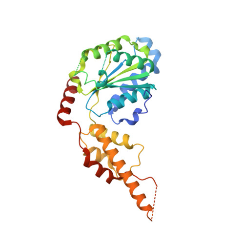
Crystal structure of the catalytic D2 domain of the AAA+ ATPase p97 reveals a putative helical split-washer-type mechanism for substrate unfolding.

Stach, L.Morgan, R.M.Makhlouf, L.Douangamath, A.von Delft, F.Zhang, X.Freemont, P.S.

(2020) FEBS Lett 594: 933-943

  • DOI: https://doi.org/10.1002/1873-3468.13667
  • Primary Citation of Related Structures:  
    6G2V, 6G2W, 6G2X, 6G2Y, 6G2Z, 6G30

  • PubMed Abstract: 

    Several pathologies have been associated with the AAA+ ATPase p97, an enzyme essential to protein homeostasis. Heterozygous polymorphisms in p97 have been shown to cause neurological disease, while elevated proteotoxic stress in tumours has made p97 an attractive cancer chemotherapy target. The cellular processes reliant on p97 are well described. High-resolution structural models of its catalytic D2 domain, however, have proved elusive, as has the mechanism by which p97 converts the energy from ATP hydrolysis into mechanical force to unfold protein substrates. Here, we describe the high-resolution structure of the p97 D2 ATPase domain. This crystal system constitutes a valuable tool for p97 inhibitor development and identifies a potentially druggable pocket in the D2 domain. In addition, its P6 1 symmetry suggests a mechanism for substrate unfolding by p97. DATABASE: The atomic coordinates and structure factors have been deposited in the PDB database under the accession numbers 6G2V, 6G2W, 6G2X, 6G2Y, 6G2Z and 6G30.


  • Organizational Affiliation

    Section of Structural and Synthetic Biology, Department of Infectious Disease, Imperial College London, UK.


Macromolecules
Find similar proteins by:  (by identity cutoff)  |  3D Structure
Entity ID: 1
MoleculeChains Sequence LengthOrganismDetailsImage
Transitional endoplasmic reticulum ATPase303Homo sapiensMutation(s): 0 
Gene Names: VCP
EC: 3.6.4.6
UniProt & NIH Common Fund Data Resources
Find proteins for P55072 (Homo sapiens)
Explore P55072 
Go to UniProtKB:  P55072
PHAROS:  P55072
GTEx:  ENSG00000165280 
Entity Groups  
Sequence Clusters30% Identity50% Identity70% Identity90% Identity95% Identity100% Identity
UniProt GroupP55072
Sequence Annotations
Expand
  • Reference Sequence
Small Molecules
Ligands 5 Unique
IDChains Name / Formula / InChI Key2D Diagram3D Interactions
ADP
Query on ADP

Download Ideal Coordinates CCD File 
H [auth A]ADENOSINE-5'-DIPHOSPHATE
C10 H15 N5 O10 P2
XTWYTFMLZFPYCI-KQYNXXCUSA-N
ELQ (Subject of Investigation/LOI)
Query on ELQ

Download Ideal Coordinates CCD File 
J [auth A][3,4-bis(fluoranyl)phenyl]-(4-methylpiperazin-1-yl)methanone
C12 H14 F2 N2 O
ZCPNSVBDWJVJSY-UHFFFAOYSA-N
MPD
Query on MPD

Download Ideal Coordinates CCD File 
B [auth A],
C [auth A],
D [auth A],
E [auth A]
(4S)-2-METHYL-2,4-PENTANEDIOL
C6 H14 O2
SVTBMSDMJJWYQN-YFKPBYRVSA-N
DMS
Query on DMS

Download Ideal Coordinates CCD File 
F [auth A],
G [auth A]
DIMETHYL SULFOXIDE
C2 H6 O S
IAZDPXIOMUYVGZ-UHFFFAOYSA-N
NA
Query on NA

Download Ideal Coordinates CCD File 
I [auth A]SODIUM ION
Na
FKNQFGJONOIPTF-UHFFFAOYSA-N
Experimental Data & Validation

Experimental Data

  • Method: X-RAY DIFFRACTION
  • Resolution: 2.15 Å
  • R-Value Free: 0.247 
  • R-Value Work: 0.220 
  • R-Value Observed: 0.221 
  • Space Group: P 61
Unit Cell:
Length ( Å )Angle ( ˚ )
a = 94.502α = 90
b = 94.502β = 90
c = 66.266γ = 120
Software Package:
Software NamePurpose
PHENIXrefinement
XDSdata reduction
XDSdata scaling
PHASERphasing

Structure Validation

View Full Validation Report


Ligand Structure Quality Assessment 


Entry History & Funding Information

Deposition Data


Funding OrganizationLocationGrant Number
Cancer Research UKUnited Kingdom--

Revision History  (Full details and data files)

  • Version 1.0: 2019-04-10
    Type: Initial release
  • Version 1.1: 2020-04-22
    Changes: Database references
  • Version 1.2: 2024-01-17
    Changes: Data collection, Database references, Refinement description