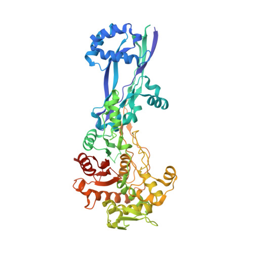
6G9F

Structural basis for the inhibition of E. coli PBP2


Experimental Data Snapshot

  • Method: X-RAY DIFFRACTION
  • Resolution: 2.35 Å
  • R-Value Free: 0.248 
  • R-Value Work: 0.199 
  • R-Value Observed: 0.201 

Starting Model: experimental
View more details

wwPDB Validation   3D Report Full Report


Ligand Structure Quality Assessment 


This is version 1.3 of the entry. See complete history


Literature

Structural Basis for E. coli Penicillin Binding Protein (PBP) 2 Inhibition, a Platform for Drug Design.

Levy, N.Bruneau, J.M.Le Rouzic, E.Bonnard, D.Le Strat, F.Caravano, A.Chevreuil, F.Barbion, J.Chasset, S.Ledoussal, B.Moreau, F.Ruff, M.

(2019) J Med Chem 62: 4742-4754

  • DOI: https://doi.org/10.1021/acs.jmedchem.9b00338
  • Primary Citation of Related Structures:  
    6G9F, 6G9P, 6G9S

  • PubMed Abstract: 

    Penicillin-binding proteins (PBPs) are the targets of the β-lactams, the most successful class of antibiotics ever developed against bacterial infections. Unfortunately, the worldwide and rapid spread of large spectrum β-lactam resistance genes such as carbapenemases is detrimental to the use of antibiotics in this class. New potent PBP inhibitors are needed, especially compounds that resist β-lactamase hydrolysis. Here we describe the structure of the E. coli PBP2 in its Apo form and upon its reaction with 2 diazabicyclo derivatives, avibactam and CPD4, a new potent PBP2 inhibitor. Examination of these structures shows that unlike avibactam, CPD4 can perform a hydrophobic stacking on Trp370 in the active site of E. coli PBP2. This result, together with sequence analysis, homology modeling, and SAR, allows us to propose CPD4 as potential starting scaffold to develop molecules active against a broad range of bacterial species at the top of the WHO priority list.


  • Organizational Affiliation

    Mutabilis , 102 Avenue Gaston Roussel , 93230 Romainville , France.


Macromolecules
Find similar proteins by:  (by identity cutoff)  |  3D Structure
Entity ID: 1
MoleculeChains Sequence LengthOrganismDetailsImage
Peptidoglycan D,D-transpeptidase MrdA561Escherichia coli K-12Mutation(s): 0 
Gene Names: mrdApbpAb0635JW0630
EC: 3.4.16.4
UniProt
Find proteins for P0AD65 (Escherichia coli (strain K12))
Explore P0AD65 
Go to UniProtKB:  P0AD65
Entity Groups  
Sequence Clusters30% Identity50% Identity70% Identity90% Identity95% Identity100% Identity
UniProt GroupP0AD65
Sequence Annotations
Expand
  • Reference Sequence
Small Molecules
Ligands 1 Unique
IDChains Name / Formula / InChI Key2D Diagram3D Interactions
NXL
Query on NXL

Download Ideal Coordinates CCD File 
B [auth A](2S,5R)-1-formyl-5-[(sulfooxy)amino]piperidine-2-carboxamide
C7 H13 N3 O6 S
WJDGWXPPFHLLNL-RITPCOANSA-N
Experimental Data & Validation

Experimental Data

  • Method: X-RAY DIFFRACTION
  • Resolution: 2.35 Å
  • R-Value Free: 0.248 
  • R-Value Work: 0.199 
  • R-Value Observed: 0.201 
  • Space Group: C 2 2 21
Unit Cell:
Length ( Å )Angle ( ˚ )
a = 125.078α = 90
b = 182.875β = 90
c = 74.767γ = 90
Software Package:
Software NamePurpose
PHENIXrefinement
XDSdata reduction
XDSdata scaling
PHENIXphasing

Structure Validation

View Full Validation Report


Ligand Structure Quality Assessment 


Entry History 

Deposition Data

Revision History  (Full details and data files)

  • Version 1.0: 2019-05-22
    Type: Initial release
  • Version 1.1: 2020-02-05
    Changes: Database references, Source and taxonomy
  • Version 1.2: 2024-01-17
    Changes: Data collection, Database references, Refinement description, Structure summary
  • Version 1.3: 2024-11-13
    Changes: Structure summary