6GG6

Crystal structure of M2 PYK in complex with Serine.


Experimental Data Snapshot

  • Method: X-RAY DIFFRACTION
  • Resolution: 2.96 Å
  • R-Value Free: 0.252 
  • R-Value Work: 0.236 
  • R-Value Observed: 0.237 

wwPDB Validation   3D Report Full Report


This is version 1.2 of the entry. See complete history


Literature

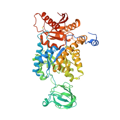
An allostatic mechanism for M2 pyruvate kinase as an amino-acid sensor.

Yuan, M.McNae, I.W.Chen, Y.Blackburn, E.A.Wear, M.A.Michels, P.A.M.Fothergill-Gilmore, L.A.Hupp, T.Walkinshaw, M.D.

(2018) Biochem J 475: 1821-1837

  • DOI: https://doi.org/10.1042/BCJ20180171
  • Primary Citation of Related Structures:  
    6GG3, 6GG4, 6GG5, 6GG6

  • PubMed Abstract: 

    We have tested the effect of all 20 proteinogenic amino acids on the activity of the M2 isoenzyme of pyruvate kinase (M2PYK) and show that, within physiologically relevant concentrations, phenylalanine, alanine, tryptophan, methionine, valine, and proline act as inhibitors, while histidine and serine act as activators. Size exclusion chromatography has been used to show that all amino acids, whether activators or inhibitors, stabilise the tetrameric form of M2PYK. In the absence of amino-acid ligands an apparent tetramer-monomer dissociation K d is estimated to be ∼0.9 µM with a slow dissociation rate ( t 1/2  15 min). X-ray structures of M2PYK complexes with alanine, phenylalanine, and tryptophan show the M2PYK locked in an inactive T-state conformation, while activators lock the M2PYK tetramer in the active R-state conformation. Amino-acid binding in the allosteric pocket triggers rigid body rotations (11°) stabilising either T or R states. The opposing inhibitory and activating effects of the non-essential amino acids serine and alanine suggest that M2PYK could act as a rapid-response nutrient sensor to rebalance cellular metabolism. This competition at a single allosteric site between activators and inhibitors provides a novel regulatory mechanism by which M2PYK activity is finely tuned by the relative (but not absolute) concentrations of activator and inhibitor amino acids. Such 'allostatic' regulation may be important in metabolic reprogramming and influencing cell fate.


  • Organizational Affiliation

    Centre for Translational and Chemical Biology, School of Biological Sciences, University of Edinburgh, Michael Swann Building, Max Born Crescent, Edinburgh EH9 3BF, U.K.


Macromolecules
Find similar proteins by:  (by identity cutoff)  |  3D Structure
Entity ID: 1
MoleculeChains Sequence LengthOrganismDetailsImage
Pyruvate kinase PKM
A, B, C, D, E
A, B, C, D, E, F, G, H
551Homo sapiensMutation(s): 0 
Gene Names: PKMOIP3PK2PK3PKM2
EC: 2.7.1.40
UniProt & NIH Common Fund Data Resources
Find proteins for P14618 (Homo sapiens)
Explore P14618 
Go to UniProtKB:  P14618
PHAROS:  P14618
GTEx:  ENSG00000067225 
Entity Groups  
Sequence Clusters30% Identity50% Identity70% Identity90% Identity95% Identity100% Identity
UniProt GroupP14618
Sequence Annotations
Expand
  • Reference Sequence
Small Molecules
Ligands 4 Unique
IDChains Name / Formula / InChI Key2D Diagram3D Interactions
SER
Query on SER

Download Ideal Coordinates CCD File 
AA [auth E]
EA [auth F]
IA [auth G]
K [auth A]
MA [auth H]
AA [auth E],
EA [auth F],
IA [auth G],
K [auth A],
MA [auth H],
O [auth B],
S [auth C],
W [auth D]
SERINE
C3 H7 N O3
MTCFGRXMJLQNBG-REOHCLBHSA-N
PO4
Query on PO4

Download Ideal Coordinates CCD File 
BA [auth E]
FA [auth F]
JA [auth G]
L [auth A]
NA [auth H]
BA [auth E],
FA [auth F],
JA [auth G],
L [auth A],
NA [auth H],
P [auth B],
T [auth C],
X [auth D]
PHOSPHATE ION
O4 P
NBIIXXVUZAFLBC-UHFFFAOYSA-K
K
Query on K

Download Ideal Coordinates CCD File 
CA [auth F]
GA [auth G]
I [auth A]
KA [auth H]
M [auth B]
CA [auth F],
GA [auth G],
I [auth A],
KA [auth H],
M [auth B],
Q [auth C],
U [auth D],
Y [auth E]
POTASSIUM ION
K
NPYPAHLBTDXSSS-UHFFFAOYSA-N
MG
Query on MG

Download Ideal Coordinates CCD File 
DA [auth F]
HA [auth G]
J [auth A]
LA [auth H]
N [auth B]
DA [auth F],
HA [auth G],
J [auth A],
LA [auth H],
N [auth B],
R [auth C],
V [auth D],
Z [auth E]
MAGNESIUM ION
Mg
JLVVSXFLKOJNIY-UHFFFAOYSA-N
Modified Residues  1 Unique
IDChains TypeFormula2D DiagramParent
CSO
Query on CSO
A, B, C, D, E
A, B, C, D, E, F, G, H
L-PEPTIDE LINKINGC3 H7 N O3 SCYS
Experimental Data & Validation

Experimental Data

  • Method: X-RAY DIFFRACTION
  • Resolution: 2.96 Å
  • R-Value Free: 0.252 
  • R-Value Work: 0.236 
  • R-Value Observed: 0.237 
  • Space Group: P 1
Unit Cell:
Length ( Å )Angle ( ˚ )
a = 93.35α = 89.72
b = 108.94β = 71.13
c = 124.34γ = 66.94
Software Package:
Software NamePurpose
REFMACrefinement
XDSdata reduction
Aimlessdata scaling
PHASERphasing

Structure Validation

View Full Validation Report


Entry History 

Deposition Data

Revision History  (Full details and data files)

  • Version 1.0: 2018-05-23
    Type: Initial release
  • Version 1.1: 2018-06-13
    Changes: Data collection, Database references
  • Version 1.2: 2024-01-17
    Changes: Data collection, Database references, Derived calculations, Refinement description