6GHV

Structure of a DC-SIGN CRD in complex with high affinity glycomimetic.


Experimental Data Snapshot

  • Method: X-RAY DIFFRACTION
  • Resolution: 2.10 Å
  • R-Value Free: 0.234 
  • R-Value Work: 0.176 
  • R-Value Observed: 0.179 

wwPDB Validation   3D Report Full Report


Ligand Structure Quality Assessment 


This is version 1.2 of the entry. See complete history


Literature

Enhancing Potency and Selectivity of a DC-SIGN Glycomimetic Ligand by Fragment-Based Design: Structural Basis.

Medve, L.Achilli, S.Guzman-Caldentey, J.Thepaut, M.Senaldi, L.Le Roy, A.Sattin, S.Ebel, C.Vives, C.Martin-Santamaria, S.Bernardi, A.Fieschi, F.

(2019) Chemistry 25: 14659-14668

  • DOI: https://doi.org/10.1002/chem.201903391
  • Primary Citation of Related Structures:  
    6GHV

  • PubMed Abstract: 

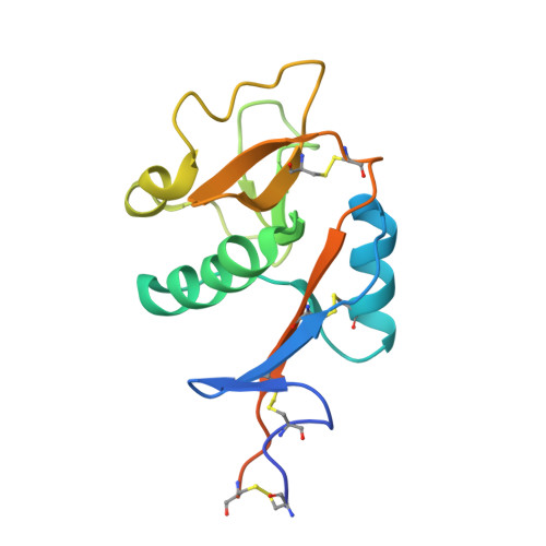
    Chemical modification of pseudo-dimannoside ligands guided by fragment-based design allowed for the exploitation of an ammonium-binding region in the vicinity of the mannose-binding site of DC-SIGN, leading to the synthesis of a glycomimetic antagonist (compound 16) of unprecedented affinity and selectivity against the related lectin langerin. Here, the computational design of pseudo-dimannoside derivatives as DC-SIGN ligands, their synthesis, their evaluation as DC-SIGN selective antagonists, the biophysical characterization of the DC-SIGN/16 complex, and the structural basis for the ligand activity are presented. On the way to the characterization of this ligand, an unusual bridging interaction within the crystals shed light on the plasticity and potential secondary binding sites within the DC-SIGN carbohydrate recognition domain.


  • Organizational Affiliation

    Dipartimento di Chimica, Università degli Studi di Milano, via Golgi 19, 20133, Milano, Italy.


Macromolecules
Find similar proteins by:  (by identity cutoff)  |  3D Structure
Entity ID: 1
MoleculeChains Sequence LengthOrganismDetailsImage
CD209 antigen
A, B, C, D, E
A, B, C, D, E, F
155Homo sapiensMutation(s): 0 
Gene Names: CD209CLEC4L
UniProt & NIH Common Fund Data Resources
Find proteins for Q9NNX6 (Homo sapiens)
Explore Q9NNX6 
Go to UniProtKB:  Q9NNX6
PHAROS:  Q9NNX6
GTEx:  ENSG00000090659 
Entity Groups  
Sequence Clusters30% Identity50% Identity70% Identity90% Identity95% Identity100% Identity
UniProt GroupQ9NNX6
Sequence Annotations
Expand
  • Reference Sequence
Small Molecules
Ligands 3 Unique
IDChains Name / Formula / InChI Key2D Diagram3D Interactions
EZ8 (Subject of Investigation/LOI)
Query on EZ8

Download Ideal Coordinates CCD File 
AA [auth D]
G [auth A]
HA [auth E]
N [auth B]
NA [auth F]
AA [auth D],
G [auth A],
HA [auth E],
N [auth B],
NA [auth F],
T [auth C]
[1-[(2~{S},3~{S},4~{R},5~{S},6~{R})-2-[(1~{S},2~{S},4~{S},5~{S})-2-(2-chloroethyloxy)-4,5-bis[[4-(hydroxymethyl)phenyl]methylcarbamoyl]cyclohexyl]oxy-6-(hydroxymethyl)-4,5-bis(oxidanyl)oxan-3-yl]-1,2,3-triazol-4-yl]methylazanium
C35 H48 Cl N6 O10
BYGZQFCUFRSCML-RWSQSUCKSA-O
CA
Query on CA

Download Ideal Coordinates CCD File 
BA [auth D]
CA [auth D]
DA [auth D]
H [auth A]
I [auth A]
BA [auth D],
CA [auth D],
DA [auth D],
H [auth A],
I [auth A],
IA [auth E],
J [auth A],
JA [auth E],
KA [auth E],
O [auth B],
OA [auth F],
P [auth B],
PA [auth F],
Q [auth B],
QA [auth F],
U [auth C],
V [auth C],
W [auth C]
CALCIUM ION
Ca
BHPQYMZQTOCNFJ-UHFFFAOYSA-N
CL
Query on CL

Download Ideal Coordinates CCD File 
EA [auth D]
FA [auth E]
GA [auth E]
K [auth A]
L [auth A]
EA [auth D],
FA [auth E],
GA [auth E],
K [auth A],
L [auth A],
LA [auth E],
M [auth A],
MA [auth F],
R [auth B],
RA [auth F],
S [auth B],
X [auth C],
Y [auth C],
Z [auth D]
CHLORIDE ION
Cl
VEXZGXHMUGYJMC-UHFFFAOYSA-M
Binding Affinity Annotations 
IDSourceBinding Affinity
EZ8 Binding MOAD:  6GHV Kd: 5.20e+4 (nM) from 1 assay(s)
Experimental Data & Validation

Experimental Data

  • Method: X-RAY DIFFRACTION
  • Resolution: 2.10 Å
  • R-Value Free: 0.234 
  • R-Value Work: 0.176 
  • R-Value Observed: 0.179 
  • Space Group: P 1 21 1
Unit Cell:
Length ( Å )Angle ( ˚ )
a = 105.612α = 90
b = 57.507β = 118.67
c = 107.247γ = 90
Software Package:
Software NamePurpose
REFMACrefinement
XDSdata reduction
XSCALEdata scaling
MOLREPphasing
PDB_EXTRACTdata extraction

Structure Validation

View Full Validation Report


Ligand Structure Quality Assessment 


Entry History & Funding Information

Deposition Data


Funding OrganizationLocationGrant Number
European CommissionFranceITN 642870

Revision History  (Full details and data files)

  • Version 1.0: 2019-09-11
    Type: Initial release
  • Version 1.1: 2019-11-27
    Changes: Database references
  • Version 1.2: 2024-01-17
    Changes: Data collection, Database references, Derived calculations, Refinement description