Quantitation of ERK1/2 inhibitor cellular target occupancies with a reversible slow off-rate probe.
Lebraud, H., Surova, O., Courtin, A., O'Reilly, M., Valenzano, C.R., Nordlund, P., Heightman, T.D.(2018) Chem Sci 9: 8608-8618
- PubMed: 30568786 Search on PubMedSearch on PubMed Central
- DOI: https://doi.org/10.1039/c8sc02754d
- Primary Citation Related Structures:
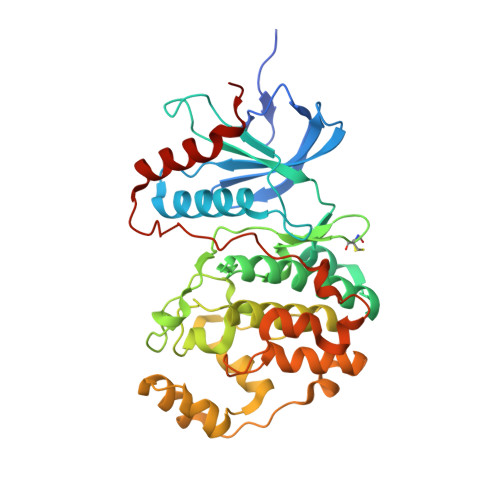
6GJB, 6GJD - PubMed Abstract:
Target engagement is a key concept in drug discovery and its direct measurement can provide a quantitative understanding of drug efficacy and/or toxicity. Failure to demonstrate target occupancy in relevant cells and tissues has been recognised as a contributing factor to the low success rate of clinical drug development. Several techniques are emerging to quantify target engagement in cells; however, in situ measurements remain challenging, mainly due to technical limitations. Here, we report the development of a non-covalent clickable probe, based on SCH772984, a slow off-rate ERK1/2 inhibitor, which enabled efficient pull down of ERK1/2 protein via click reaction with tetrazine tagged agarose beads. This was used in a competition setting to measure relative target occupancy by selected ERK1/2 inhibitors. As a reference we used the cellular thermal shift assay, a label-free biophysical assay relying solely on ligand-induced thermodynamic stabilization of proteins. To validate the EC 50 values measured by both methods, the results were compared with IC 50 data for the phosphorylation of RSK, a downstream substrate of ERK1/2 used as a functional biomarker of ERK1/2 inhibition. We showed that a slow off-rate reversible probe can be used to efficiently pull down cellular proteins, significantly extending the potential of the approach beyond the need for covalent or photoaffinity warheads.
- Astex Pharmaceuticals , 436 Cambridge Science Park , Cambridge , CB4 0QA , UK . Email: honorine.lebraud@astx.com ; Email: tom.heightman@astx.com.
Organizational Affiliation: