6GJH | pdb_00006gjh

Human Hsp27 (HspB1) alpha-crystallin domain in complex with a peptide mimic of its phosphorylatable N-terminal region


Experimental Data Snapshot

  • Method: X-RAY DIFFRACTION
  • Resolution: 2.10 Å
  • R-Value Free: 
    0.251 (Depositor), 0.215 (DCC) 
  • R-Value Work: 
    0.211 (Depositor), 0.218 (DCC) 
  • R-Value Observed: 
    0.213 (Depositor) 

Starting Model: experimental
View more details

wwPDB Validation 3D Report Full Report

Validation slider image for 6GJH

This is version 1.3 of the entry. See complete history

Literature

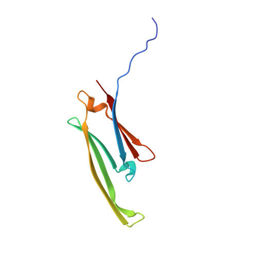
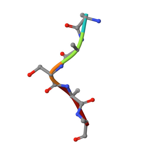
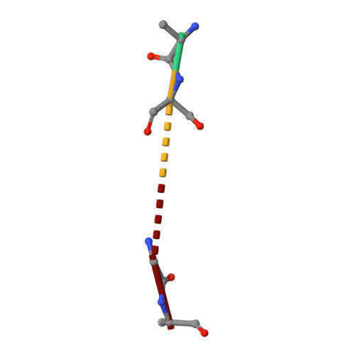
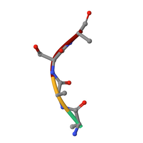
HspB1 phosphorylation regulates its intramolecular dynamics and mechanosensitive molecular chaperone interaction with filamin C.

Collier, M.P.Alderson, T.R.de Villiers, C.P.Nicholls, D.Gastall, H.Y.Allison, T.M.Degiacomi, M.T.Jiang, H.Mlynek, G.Furst, D.O.van der Ven, P.F.M.Djinovic-Carugo, K.Baldwin, A.J.Watkins, H.Gehmlich, K.Benesch, J.L.P.

(2019) Sci Adv 5: eaav8421-eaav8421

  • DOI: https://doi.org/10.1126/sciadv.aav8421
  • Primary Citation Related Structures: 
    6GJH

  • PubMed Abstract: 

    Mechanical force-induced conformational changes in proteins underpin a variety of physiological functions, typified in muscle contractile machinery. Mutations in the actin-binding protein filamin C (FLNC) are linked to musculoskeletal pathologies characterized by altered biomechanical properties and sometimes aggregates. HspB1, an abundant molecular chaperone, is prevalent in striated muscle where it is phosphorylated in response to cues including mechanical stress. We report the interaction and up-regulation of both proteins in three mouse models of biomechanical stress, with HspB1 being phosphorylated and FLNC being localized to load-bearing sites. We show how phosphorylation leads to increased exposure of the residues surrounding the HspB1 phosphosite, facilitating their binding to a compact multidomain region of FLNC proposed to have mechanosensing functions. Steered unfolding of FLNC reveals that its extension trajectory is modulated by the phosphorylated region of HspB1. This may represent a posttranslationally regulated chaperone-client protection mechanism targeting over-extension during mechanical stress.


  • Organizational Affiliation
    • Department of Chemistry, Physical and Theoretical Chemistry Laboratory, University of Oxford, South Parks Road, Oxford OX1 3QZ, UK.

Macromolecule Content 

  • Total Structure Weight: 79.68 kDa 
  • Atom Count: 6,007 
  • Modeled Residue Count: 712 
  • Deposited Residue Count: 713 
  • Unique protein chains: 4

Macromolecules

Find similar proteins by:|  3D Structure
Entity ID: 1
MoleculeChains  Sequence LengthOrganismDetailsImage
Heat shock protein beta-187Homo sapiensMutation(s): 0 
Gene Names: HSPB1HSP27HSP28
UniProt & NIH Common Fund Data Resources
Find proteins for P04792 (Homo sapiens)
Explore P04792 
Go to UniProtKB:  P04792
PHAROS:  P04792
GTEx:  ENSG00000106211 
Entity Groups
Sequence Clusters30% Identity50% Identity70% Identity90% Identity95% Identity100% Identity
UniProt GroupP04792
Sequence Annotations
Expand
Reference Sequence
Find similar proteins by:  Sequence   |   3D Structure  
Entity ID: 2
MoleculeChains  Sequence LengthOrganismDetailsImage
ALA-LEU-SER-ARG-GLNI [auth K]5Homo sapiensMutation(s): 0 
Find similar proteins by:  Sequence   |   3D Structure  
Entity ID: 3
MoleculeChains  Sequence LengthOrganismDetailsImage
LEU-SER-GLY-VAL4Homo sapiensMutation(s): 0 
Find similar proteins by:  Sequence   |   3D Structure  
Entity ID: 4
MoleculeChains  Sequence LengthOrganismDetailsImage
ALA-LEU-SER-ARGK [auth L],
L [auth I]
4Homo sapiensMutation(s): 0 

Small Molecules

Ligands 1 Unique
IDChains Name / Formula / InChI Key2D Diagram3D Interactions
GOL

Query on GOL


Download:Ideal Coordinates CCD File
M [auth A]GLYCEROL
C3 H8 O3
PEDCQBHIVMGVHV-UHFFFAOYSA-N

Experimental Data & Validation

Experimental Data

  • Method: X-RAY DIFFRACTION
  • Resolution: 2.10 Å
  • R-Value Free:  0.251 (Depositor), 0.215 (DCC) 
  • R-Value Work:  0.211 (Depositor), 0.218 (DCC) 
  • R-Value Observed: 0.213 (Depositor) 
Space Group: P 1 21 1
Unit Cell:
Length ( Å )Angle ( ˚ )
a = 56.81α = 90
b = 157.67β = 95.45
c = 57.87γ = 90
Software Package:
Software NamePurpose
PHENIXrefinement
MOSFLMdata reduction
Aimlessdata scaling
PHASERphasing
PDB_EXTRACTdata extraction

Structure Validation

View Full Validation Report


Entry History 

& Funding Information

Deposition Data


Funding OrganizationLocationGrant Number
Biotechnology and Biological Sciences Research CouncilUnited KingdomBB/K004247/1
Biotechnology and Biological Sciences Research CouncilUnited KingdomBB/J018082/1

Revision History  (Full details and data files)

  • Version 1.0: 2019-05-29
    Type: Initial release
  • Version 1.1: 2019-06-12
    Changes: Data collection, Database references
  • Version 1.2: 2024-01-17
    Changes: Data collection, Database references, Refinement description
  • Version 1.3: 2024-10-16
    Changes: Structure summary