6GMD

The crystal structure of CK2alpha in complex with compound 3


Experimental Data Snapshot

  • Method: X-RAY DIFFRACTION
  • Resolution: 1.66 Å
  • R-Value Free: 0.247 
  • R-Value Work: 0.221 
  • R-Value Observed: 0.222 

wwPDB Validation   3D Report Full Report


Ligand Structure Quality Assessment 


This is version 1.2 of the entry. See complete history


Literature

Novel non-ATP competitive small molecules targeting the CK2 alpha / beta interface.

Brear, P.North, A.Iegre, J.Hadje Georgiou, K.Lubin, A.Carro, L.Green, W.Sore, H.F.Hyvonen, M.Spring, D.R.

(2018) Bioorg Med Chem 26: 3016-3020

  • DOI: https://doi.org/10.1016/j.bmc.2018.05.011
  • Primary Citation of Related Structures:  
    6GIH, 6GMD

  • PubMed Abstract: 

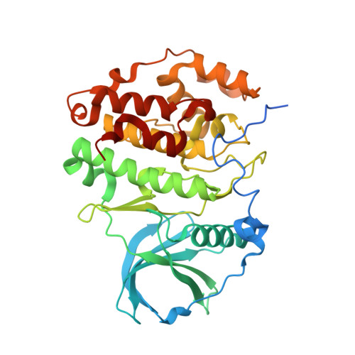
    Increased CK2 levels are prevalent in many cancers. Combined with the critical role CK2 plays in many cell-signaling pathways, this makes it a prime target for down regulation to fight tumour growth. Herein, we report a fragment-based approach to inhibiting the interaction between CK2α and CK2β at the α-β interface of the holoenzyme. A fragment, CAM187, with an IC 50 of 44 μM and a molecular weight of only 257 gmol -1 has been identified as the most promising compound. Importantly, the lead fragment only bound at the interface and was not observed in the ATP binding site of the protein when co-crystallised with CK2α. The fragment-like molecules discovered in this study represent unique scaffolds to CK2 inhibition and leave room for further optimisation.


  • Organizational Affiliation

    Department of Biochemistry, University of Cambridge, Sanger Building, 80 Tennis Court Road, Old Addenbrooke's Site, Cambridge CB2 1GA, UK.


Macromolecules
Find similar proteins by:  (by identity cutoff)  |  3D Structure
Entity ID: 1
MoleculeChains Sequence LengthOrganismDetailsImage
Casein kinase II subunit alpha
A, B
352Homo sapiensMutation(s): 1 
Gene Names: CSNK2A1CK2A1
EC: 2.7.11.1
UniProt & NIH Common Fund Data Resources
Find proteins for P68400 (Homo sapiens)
Explore P68400 
Go to UniProtKB:  P68400
PHAROS:  P68400
GTEx:  ENSG00000101266 
Entity Groups  
Sequence Clusters30% Identity50% Identity70% Identity90% Identity95% Identity100% Identity
UniProt GroupP68400
Sequence Annotations
Expand
  • Reference Sequence
Small Molecules
Ligands 3 Unique
IDChains Name / Formula / InChI Key2D Diagram3D Interactions
ATP
Query on ATP

Download Ideal Coordinates CCD File 
E [auth A],
K [auth B]
ADENOSINE-5'-TRIPHOSPHATE
C10 H16 N5 O13 P3
ZKHQWZAMYRWXGA-KQYNXXCUSA-N
A8Q
Query on A8Q

Download Ideal Coordinates CCD File 
D [auth A],
I [auth B],
J [auth B]
[3-chloranyl-4-(2-propan-2-ylphenyl)phenyl]methylazanium
C16 H19 Cl N
XAFZSQVXSGXDQY-UHFFFAOYSA-O
ACT
Query on ACT

Download Ideal Coordinates CCD File 
C [auth A],
F [auth B],
G [auth B],
H [auth B]
ACETATE ION
C2 H3 O2
QTBSBXVTEAMEQO-UHFFFAOYSA-M
Experimental Data & Validation

Experimental Data

  • Method: X-RAY DIFFRACTION
  • Resolution: 1.66 Å
  • R-Value Free: 0.247 
  • R-Value Work: 0.221 
  • R-Value Observed: 0.222 
  • Space Group: C 2 2 21
Unit Cell:
Length ( Å )Angle ( ˚ )
a = 64.739α = 90
b = 68.84β = 90
c = 333.258γ = 90
Software Package:
Software NamePurpose
BUSTERrefinement
Aimlessdata scaling
PDB_EXTRACTdata extraction
DIALSdata reduction
PHASERphasing

Structure Validation

View Full Validation Report


Ligand Structure Quality Assessment 


Entry History & Funding Information

Deposition Data


Funding OrganizationLocationGrant Number
Wellcome TrustUnited Kingdom090340/Z/09/Z
Wellcome TrustUnited Kingdom107714/Z/15/Z

Revision History  (Full details and data files)

  • Version 1.0: 2018-06-06
    Type: Initial release
  • Version 1.1: 2018-06-20
    Changes: Data collection, Database references
  • Version 1.2: 2024-01-17
    Changes: Data collection, Database references, Refinement description