An Unusual Intramolecular Halogen Bond Guides Conformational Selection.
Tesch, R., Becker, C., Muller, M.P., Beck, M.E., Quambusch, L., Getlik, M., Lategahn, J., Uhlenbrock, N., Costa, F.N., Poleto, M.D., Pinheiro, P.S.M., Rodrigues, D.A., Sant'Anna, C.M.R., Ferreira, F.F., Verli, H., Fraga, C.A.M., Rauh, D.(2018) Angew Chem Int Ed Engl 57: 9970-9975
- PubMed: 29873877 Search on PubMed
- DOI: https://doi.org/10.1002/anie.201804917
- Primary Citation Related Structures:
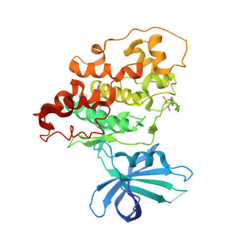
6GN1 - PubMed Abstract:
PIK-75 is a phosphoinositide-3-kinase (PI3K) α-isoform-selective inhibitor with high potency. Although published structure-activity relationship data show the importance of the NO 2 and the Br substituents in PIK-75, none of the published studies could correctly determine the underlying reason for their importance. In this publication, we report the first X-ray crystal structure of PIK-75 in complex with the kinase GSK-3β. The structure shows an unusual U-shaped conformation of PIK-75 within the active site of GSK-3β that is likely stabilized by an atypical intramolecular Br⋅⋅⋅NO 2 halogen bond. NMR and MD simulations show that this conformation presumably also exists in solution and leads to a binding-competent preorganization of the PIK-75 molecule, thus explaining its high potency. We therefore suggest that the site-specific incorporation of halogen bonds could be generally used to design conformationally restricted bioactive substances with increased potencies.
- Faculty of Chemistry and Chemical Biology, TU Dortmund University, Otto-Hahn-Strasse 4a, 44227, Dortmund, Germany.
Organizational Affiliation: