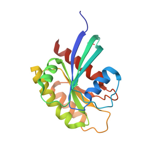
6GOM

KRAS-169 Q61H GPPNHP + PPIN-1


Experimental Data Snapshot

  • Method: X-RAY DIFFRACTION
  • Resolution: 1.63 Å
  • R-Value Free: 0.207 
  • R-Value Work: 0.186 
  • R-Value Observed: 0.187 

Starting Model: experimental
View more details

wwPDB Validation   3D Report Full Report


Ligand Structure Quality Assessment 


This is version 1.3 of the entry. See complete history


Literature

Structure-based development of new RAS-effector inhibitors from a combination of active and inactive RAS-binding compounds.

Cruz-Migoni, A.Canning, P.Quevedo, C.E.Bataille, C.J.R.Bery, N.Miller, A.Russell, A.J.Phillips, S.E.V.Carr, S.B.Rabbitts, T.H.

(2019) Proc Natl Acad Sci U S A 116: 2545-2550

  • DOI: https://doi.org/10.1073/pnas.1811360116
  • Primary Citation of Related Structures:  
    6GOD, 6GOE, 6GOF, 6GOG, 6GOM, 6GQT, 6GQW, 6GQX, 6GQY

  • PubMed Abstract: 

    The RAS gene family is frequently mutated in human cancers, and the quest for compounds that bind to mutant RAS remains a major goal, as it also does for inhibitors of protein-protein interactions. We have refined crystallization conditions for KRAS 169 Q61H -yielding crystals suitable for soaking with compounds and exploited this to assess new RAS-binding compounds selected by screening a protein-protein interaction-focused compound library using surface plasmon resonance. Two compounds, referred to as PPIN-1 and PPIN-2, with related structures from 30 initial RAS binders showed binding to a pocket where compounds had been previously developed, including RAS effector protein-protein interaction inhibitors selected using an intracellular antibody fragment (called Abd compounds). Unlike the Abd series of RAS binders, PPIN-1 and PPIN-2 compounds were not competed by the inhibitory anti-RAS intracellular antibody fragment and did not show any RAS-effector inhibition properties. By fusing the common, anchoring part from the two new compounds with the inhibitory substituents of the Abd series, we have created a set of compounds that inhibit RAS-effector interactions with increased potency. These fused compounds add to the growing catalog of RAS protein-protein inhibitors and show that building a chemical series by crossing over two chemical series is a strategy to create RAS-binding small molecules.


  • Organizational Affiliation

    Weatherall Institute of Molecular Medicine, MRC Molecular Haematology Unit, John Radcliffe Hospital, University of Oxford, OX3 9DS Oxford, United Kingdom.


Macromolecules
Find similar proteins by:  (by identity cutoff)  |  3D Structure
Entity ID: 1
MoleculeChains Sequence LengthOrganismDetailsImage
GTPase KRas
A, B, C, D, E
A, B, C, D, E, F
172Homo sapiensMutation(s): 1 
Gene Names: KRASKRAS2RASK2
EC: 3.6.5.2
UniProt & NIH Common Fund Data Resources
Find proteins for P01116 (Homo sapiens)
Explore P01116 
Go to UniProtKB:  P01116
PHAROS:  P01116
GTEx:  ENSG00000133703 
Entity Groups  
Sequence Clusters30% Identity50% Identity70% Identity90% Identity95% Identity100% Identity
UniProt GroupP01116
Sequence Annotations
Expand
  • Reference Sequence
Small Molecules
Ligands 5 Unique
IDChains Name / Formula / InChI Key2D Diagram3D Interactions
GNP
Query on GNP

Download Ideal Coordinates CCD File 
G [auth A]
L [auth B]
O [auth C]
Q [auth D]
T [auth E]
G [auth A],
L [auth B],
O [auth C],
Q [auth D],
T [auth E],
V [auth F]
PHOSPHOAMINOPHOSPHONIC ACID-GUANYLATE ESTER
C10 H17 N6 O13 P3
UQABYHGXWYXDTK-UUOKFMHZSA-N
F6E
Query on F6E

Download Ideal Coordinates CCD File 
J [auth A](6~{S})-1-(1~{H}-imidazol-4-ylcarbonyl)-6-[(4-phenylphenyl)methyl]-4-propyl-1,4-diazepan-5-one
C25 H28 N4 O2
NXIUWTFGGYLRTA-QFIPXVFZSA-N
CIT
Query on CIT

Download Ideal Coordinates CCD File 
I [auth A],
K [auth A],
S [auth D]
CITRIC ACID
C6 H8 O7
KRKNYBCHXYNGOX-UHFFFAOYSA-N
PO4
Query on PO4

Download Ideal Coordinates CCD File 
N [auth B]PHOSPHATE ION
O4 P
NBIIXXVUZAFLBC-UHFFFAOYSA-K
MG
Query on MG

Download Ideal Coordinates CCD File 
H [auth A]
M [auth B]
P [auth C]
R [auth D]
U [auth E]
H [auth A],
M [auth B],
P [auth C],
R [auth D],
U [auth E],
W [auth F]
MAGNESIUM ION
Mg
JLVVSXFLKOJNIY-UHFFFAOYSA-N
Modified Residues  1 Unique
IDChains TypeFormula2D DiagramParent
CSO
Query on CSO
A, B, C, D, E
A, B, C, D, E, F
L-PEPTIDE LINKINGC3 H7 N O3 SCYS
Experimental Data & Validation

Experimental Data

  • Method: X-RAY DIFFRACTION
  • Resolution: 1.63 Å
  • R-Value Free: 0.207 
  • R-Value Work: 0.186 
  • R-Value Observed: 0.187 
  • Space Group: P 21 21 21
Unit Cell:
Length ( Å )Angle ( ˚ )
a = 64.18α = 90
b = 119.84β = 90
c = 158.6γ = 90
Software Package:
Software NamePurpose
REFMACrefinement
xia2data reduction
xia2data scaling
PHASERphasing

Structure Validation

View Full Validation Report


Ligand Structure Quality Assessment 


Entry History & Funding Information

Deposition Data


Funding OrganizationLocationGrant Number
Wellcome TrustUnited Kingdom099246/Z/12/Z
BloodwiseUnited Kingdom12051
Medical Research Council (United Kingdom)United KingdomMR/J000612/1

Revision History  (Full details and data files)

  • Version 1.0: 2019-02-06
    Type: Initial release
  • Version 1.1: 2019-02-20
    Changes: Data collection, Database references
  • Version 1.2: 2024-01-17
    Changes: Data collection, Database references, Derived calculations, Refinement description
  • Version 1.3: 2024-11-06
    Changes: Structure summary