6GOT | pdb_00006got

Crystal structure of human carbonic anhydrase II in complex with the inhibitor 4-(phenethylthio)benzenesulfonamide


Experimental Data Snapshot

  • Method: X-RAY DIFFRACTION
  • Resolution: 1.56 Å
  • R-Value Free: 
    0.223 (Depositor), 0.223 (DCC) 
  • R-Value Work: 
    0.199 (Depositor), 0.199 (DCC) 
  • R-Value Observed: 
    0.200 (Depositor) 

Starting Model: experimental
View more details

wwPDB Validation 3D Report Full Report

Validation slider image for 6GOT

Ligand Structure Quality Assessment 


This is version 1.2 of the entry. See complete history

Literature

Synthesis of different thio-scaffolds bearing sulfonamide with subnanomolar carbonic anhydrase II and IX inhibitory properties and X-ray investigations for their inhibitory mechanism.

Angeli, A.Tanini, D.Capperucci, A.Malevolti, G.Turco, F.Ferraroni, M.Supuran, C.T.

(2018) Bioorg Chem 81: 642-648

  • DOI: https://doi.org/10.1016/j.bioorg.2018.09.028
  • Primary Citation Related Structures: 
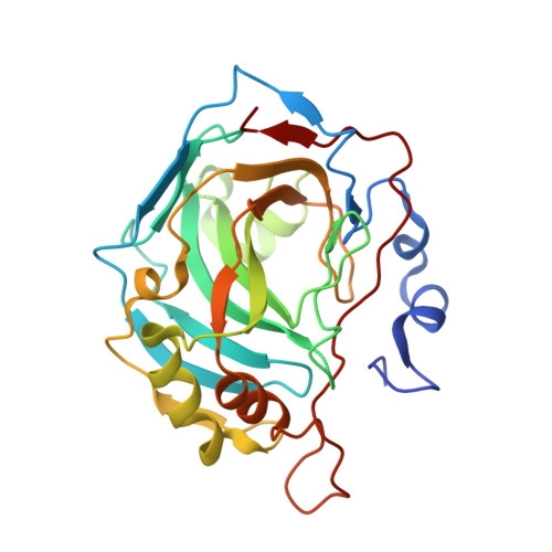
    6GOT

  • PubMed Abstract: 

    Several new molecules with different thio-scaffolds were designed, synthesised, and evaluated biologically as inhibitors of Carbonic Anhydrases (CAIs). The structure-activity relationship analysis identified thioether derivatives, here reported, as a potent and selective CAIs against hCA II and hCA IX. High resolution X-ray structure of inhibitor bound hCA II revealed extensive interactions with the hydrophobic pocket of active site and provided molecular insight into the binding properties of these new inhibitors.


  • Organizational Affiliation
    • Department of University of Florence, NEUROFARBA Dept., Sezione di Scienze Farmaceutiche, Via Ugo Schiff 6, 50019 Sesto Fiorentino (Florence), Italy.

Macromolecule Content 

  • Total Structure Weight: 29.83 kDa 
  • Atom Count: 2,218 
  • Modeled Residue Count: 257 
  • Deposited Residue Count: 260 
  • Unique protein chains: 1

Macromolecules

Find similar proteins by:|  3D Structure
Entity ID: 1
MoleculeChains  Sequence LengthOrganismDetailsImage
Carbonic anhydrase 2260Homo sapiensMutation(s): 0 
EC: 4.2.1.1 (PDB Primary Data), 4.2.1.69 (UniProt)
UniProt & NIH Common Fund Data Resources
Find proteins for P00918 (Homo sapiens)
Explore P00918 
Go to UniProtKB:  P00918
PHAROS:  P00918
GTEx:  ENSG00000104267 
Entity Groups
Sequence Clusters30% Identity50% Identity70% Identity90% Identity95% Identity100% Identity
UniProt GroupP00918
Sequence Annotations
Expand
Reference Sequence

Experimental Data & Validation

Experimental Data

  • Method: X-RAY DIFFRACTION
  • Resolution: 1.56 Å
  • R-Value Free:  0.223 (Depositor), 0.223 (DCC) 
  • R-Value Work:  0.199 (Depositor), 0.199 (DCC) 
  • R-Value Observed: 0.200 (Depositor) 
Space Group: P 1 21 1
Unit Cell:
Length ( Å )Angle ( ˚ )
a = 42.443α = 90
b = 41.284β = 104.38
c = 72.171γ = 90
Software Package:
Software NamePurpose
XDSdata reduction
XSCALEdata scaling
REFMACrefinement
PDB_EXTRACTdata extraction
REFMACphasing

Structure Validation

View Full Validation Report


Ligand Structure Quality Assessment 


Entry History 

Deposition Data

Revision History  (Full details and data files)

  • Version 1.0: 2018-10-03
    Type: Initial release
  • Version 1.1: 2018-10-10
    Changes: Data collection, Structure summary
  • Version 1.2: 2024-01-17
    Changes: Advisory, Data collection, Database references, Refinement description, Structure summary