6H22

Crystal structure of Mdm2 bound to a stapled peptide


Experimental Data Snapshot

  • Method: X-RAY DIFFRACTION
  • Resolution: 2.01 Å
  • R-Value Free: 0.238 
  • R-Value Work: 0.211 
  • R-Value Observed: 0.212 

wwPDB Validation   3D Report Full Report


Ligand Structure Quality Assessment 


This is version 1.2 of the entry. See complete history


Literature

Water-soluble, stable and azide-reactive strained dialkynes for biocompatible double strain-promoted click chemistry.

Sharma, K.Strizhak, A.V.Fowler, E.Wang, X.Xu, W.Hatt Jensen, C.Wu, Y.Sore, H.F.Lau, Y.H.Hyvonen, M.Itzhaki, L.S.Spring, D.R.

(2019) Org Biomol Chem 17: 8014-8018

  • DOI: https://doi.org/10.1039/c9ob01745c
  • Primary Citation of Related Structures:  
    6H22

  • PubMed Abstract: 

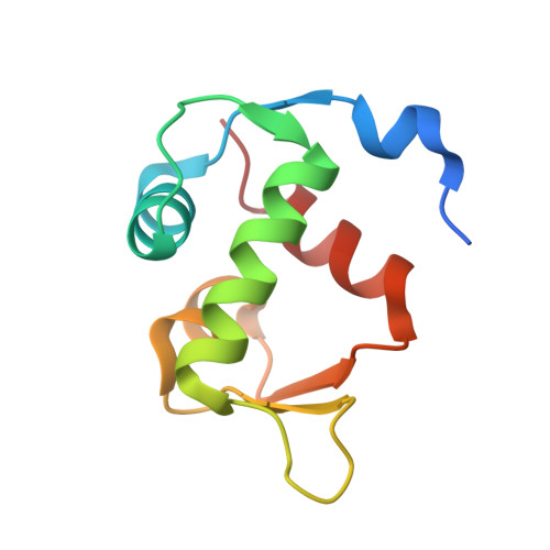
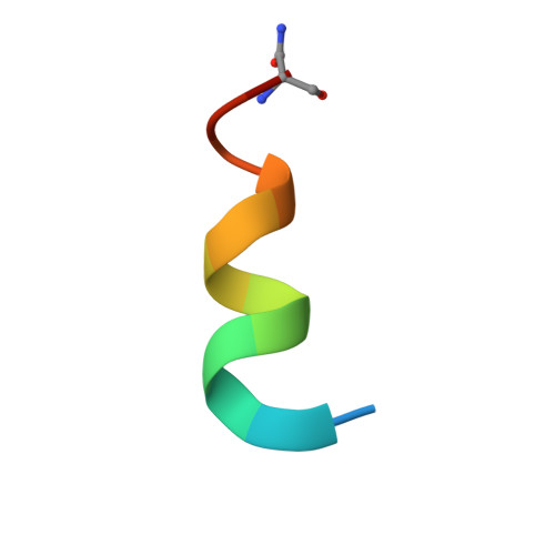
    The Sondheimer dialkyne is extensively used in double strain-promoted azide-alkyne cycloadditions. This reagent suffers with poor water-solubility and rapidly decomposes in aqueous solutions. This intrinsically limits its application in biological systems, and no effective solutions are currently available. Herein, we report the development of novel highly water-soluble, stable, and azide-reactive strained dialkyne reagents. To demonstrate their extensive utility, we applied our novel dialkynes to a double strain-promoted macrocyclisation strategy to generate functionalised p53-based stapled peptides for inhibiting the oncogenic p53-MDM2 interaction. These functionalised stapled peptides bind MDM2 with low nanomolar affinity and show p53 activation in a cellular environment. Overall, our highly soluble, stable and azide-reactive dialkynes offer significant advantages over the currently used Sondheimer dialkyne, and could be utilised for numerous biological applications.


  • Organizational Affiliation

    Department of Chemistry, University of Cambridge, Lensfield Road, Cambridge CB2 1EW, UK.


Macromolecules
Find similar proteins by:  (by identity cutoff)  |  3D Structure
Entity ID: 1
MoleculeChains Sequence LengthOrganismDetailsImage
E3 ubiquitin-protein ligase Mdm2
A, B
97Homo sapiensMutation(s): 0 
Gene Names: MDM2
EC: 2.3.2.27
UniProt & NIH Common Fund Data Resources
Find proteins for Q00987 (Homo sapiens)
Explore Q00987 
Go to UniProtKB:  Q00987
PHAROS:  Q00987
GTEx:  ENSG00000135679 
Entity Groups  
Sequence Clusters30% Identity50% Identity70% Identity90% Identity95% Identity100% Identity
UniProt GroupQ00987
Sequence Annotations
Expand
  • Reference Sequence

Find similar proteins by:  Sequence   |   3D Structure  

Entity ID: 2
MoleculeChains Sequence LengthOrganismDetailsImage
Stapled peptide
C, D
14Homo sapiensMutation(s): 0 
Entity Groups  
Sequence Clusters30% Identity50% Identity70% Identity90% Identity95% Identity100% Identity
Sequence Annotations
Expand
  • Reference Sequence
Small Molecules
Ligands 1 Unique
IDChains Name / Formula / InChI Key2D Diagram3D Interactions
FL5 (Subject of Investigation/LOI)
Query on FL5

Download Ideal Coordinates CCD File 
E [auth C],
F [auth D]
12-(dimethylamino)-3,10-diethyl-N,N,N-trimethyl-3,10-dihydrodibenzo[3,4:7,8]cycloocta[1,2-d:5,6-d']bis([1,2,3]triazole)-5-aminium
C25 H31 N8
QOPAGBBAMAJINO-MOBSCGTJSA-N
Experimental Data & Validation

Experimental Data

  • Method: X-RAY DIFFRACTION
  • Resolution: 2.01 Å
  • R-Value Free: 0.238 
  • R-Value Work: 0.211 
  • R-Value Observed: 0.212 
  • Space Group: P 21 21 21
Unit Cell:
Length ( Å )Angle ( ˚ )
a = 64.416α = 90
b = 72.186β = 90
c = 45.124γ = 90
Software Package:
Software NamePurpose
PHENIXrefinement
Aimlessdata scaling
PDB_EXTRACTdata extraction
XDSdata reduction
PHASERphasing

Structure Validation

View Full Validation Report


Ligand Structure Quality Assessment 


Entry History 

Deposition Data

Revision History  (Full details and data files)

  • Version 1.0: 2019-07-31
    Type: Initial release
  • Version 1.1: 2019-08-28
    Changes: Data collection, Database references
  • Version 1.2: 2019-09-11
    Changes: Data collection, Database references