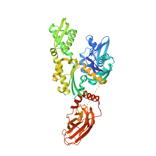
6H54

CRYSTAL STRUCTURE OF BOVINE HSC70(AA1-554)E213A/D214A IN COMPLEX WITH INHIBITOR VER155008


Experimental Data Snapshot

  • Method: X-RAY DIFFRACTION
  • Resolution: 2.02 Å
  • R-Value Free: 0.258 
  • R-Value Work: 0.221 
  • R-Value Observed: 0.222 

Starting Model: experimental
View more details

wwPDB Validation   3D Report Full Report


Ligand Structure Quality Assessment 


This is version 2.2 of the entry. See complete history


Literature

Combined In-Solution Fragment Screening and Crystallographic Binding-Mode Analysis with a Two-Domain Hsp70 Construct.

Zehe, M.Kehrein, J.Schollmayer, C.Plank, C.Kovacs, H.Merino Asumendi, E.Holzgrabe, U.Grimm, C.Sotriffer, C.

(2024) ACS Chem Biol 

  • DOI: https://doi.org/10.1021/acschembio.3c00589
  • Primary Citation of Related Structures:  
    6H54, 7O6R, 7ODB, 7ODD, 7ODI, 7PLK

  • PubMed Abstract: 

    Heat shock protein 70 (Hsp70) isoforms are key players in the regulation of protein homeostasis and cell death pathways and are therefore attractive targets in cancer research. Developing nucleotide-competitive inhibitors or allosteric modulators, however, has turned out to be very challenging for this protein family, and no Hsp70-directed therapeutics have so far become available. As the field could profit from alternative starting points for inhibitor development, we present the results of a fragment-based screening approach on a two-domain Hsp70 construct using in-solution NMR methods, together with X-ray-crystallographic investigations and mixed-solvent molecular dynamics simulations. The screening protocol resulted in hits on both domains. In particular, fragment binding in a deeply buried pocket at the substrate-binding domain could be detected. The corresponding site is known to be important for communication between the nucleotide-binding and substrate-binding domains of Hsp70 proteins. The main fragment identified at this position also offers an interesting starting point for the development of a dual Hsp70/Hsp90 inhibitor.


  • Organizational Affiliation

    University of Würzburg, Institute of Pharmacy and Food Chemistry, Am Hubland, DE-97074 Würzburg, Germany.


Macromolecules
Find similar proteins by:  (by identity cutoff)  |  3D Structure
Entity ID: 1
MoleculeChains Sequence LengthOrganismDetailsImage
Heat shock cognate 71 kDa protein554Bos taurusMutation(s): 2 
Gene Names: HSPA8HSC70
EC: 3.6.4.10
UniProt
Find proteins for P19120 (Bos taurus)
Explore P19120 
Go to UniProtKB:  P19120
Entity Groups  
Sequence Clusters30% Identity50% Identity70% Identity90% Identity95% Identity100% Identity
UniProt GroupP19120
Sequence Annotations
Expand
  • Reference Sequence
Small Molecules
Ligands 4 Unique
IDChains Name / Formula / InChI Key2D Diagram3D Interactions
3FD (Subject of Investigation/LOI)
Query on 3FD

Download Ideal Coordinates CCD File 
C [auth A]4-[[(2R,3S,4R,5R)-5-[6-amino-8-[(3,4-dichlorophenyl)methylamino]purin-9-yl]-3,4-dihydroxy-oxolan-2-yl]methoxymethyl]benzonitrile
C25 H23 Cl2 N7 O4
ZXGGCBQORXDVTE-UMCMBGNQSA-N
EPE
Query on EPE

Download Ideal Coordinates CCD File 
D [auth A]4-(2-HYDROXYETHYL)-1-PIPERAZINE ETHANESULFONIC ACID
C8 H18 N2 O4 S
JKMHFZQWWAIEOD-UHFFFAOYSA-N
GOL
Query on GOL

Download Ideal Coordinates CCD File 
E [auth A]GLYCEROL
C3 H8 O3
PEDCQBHIVMGVHV-UHFFFAOYSA-N
TMO
Query on TMO

Download Ideal Coordinates CCD File 
B [auth A]trimethylamine oxide
C3 H9 N O
UYPYRKYUKCHHIB-UHFFFAOYSA-N
Experimental Data & Validation

Experimental Data

  • Method: X-RAY DIFFRACTION
  • Resolution: 2.02 Å
  • R-Value Free: 0.258 
  • R-Value Work: 0.221 
  • R-Value Observed: 0.222 
  • Space Group: P 1 21 1
Unit Cell:
Length ( Å )Angle ( ˚ )
a = 66.19α = 90
b = 49.34β = 99.801
c = 86.96γ = 90
Software Package:
Software NamePurpose
PHENIXrefinement
PHENIXrefinement
PDB_EXTRACTdata extraction
XDSdata reduction
XDSdata scaling
PHASERphasing

Structure Validation

View Full Validation Report


Ligand Structure Quality Assessment 


Entry History & Funding Information

Deposition Data


Funding OrganizationLocationGrant Number
German Research Foundation (DFG)GermanyCRU 216

Revision History  (Full details and data files)

  • Version 1.0: 2019-08-14
    Type: Initial release
  • Version 1.1: 2019-08-21
    Changes: Data collection
  • Version 2.0: 2020-07-08
    Type: Coordinate replacement
    Reason: Ligand geometry
    Changes: Advisory, Atomic model, Author supporting evidence, Data collection, Database references, Derived calculations, Non-polymer description, Other, Refinement description, Source and taxonomy, Structure summary
  • Version 2.1: 2024-01-17
    Changes: Advisory, Data collection, Database references, Refinement description
  • Version 2.2: 2024-02-14
    Changes: Database references