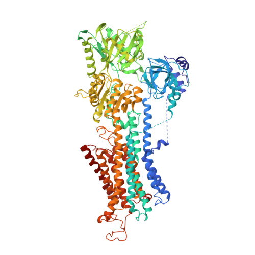
6HXB

SERCA2a from pig heart


Experimental Data Snapshot

  • Method: X-RAY DIFFRACTION
  • Resolution: 4.00 Å
  • R-Value Free: 0.354 
  • R-Value Work: 0.310 

wwPDB Validation   3D Report Full Report


Ligand Structure Quality Assessment 


This is version 1.3 of the entry. See complete history


Literature

Structures of the heart specific SERCA2a Ca 2+ -ATPase.

Sitsel, A.De Raeymaecker, J.Drachmann, N.D.Derua, R.Smaardijk, S.Andersen, J.L.Vandecaetsbeek, I.Chen, J.De Maeyer, M.Waelkens, E.Olesen, C.Vangheluwe, P.Nissen, P.

(2019) EMBO J 38

  • DOI: https://doi.org/10.15252/embj.2018100020
  • Primary Citation of Related Structures:  
    5MPM, 6HXB

  • PubMed Abstract: 

    The sarcoplasmic/endoplasmic reticulum Ca 2+ -ATPase 2a (SERCA2a) performs active reuptake of cytoplasmic Ca 2+ and is a major regulator of cardiac muscle contractility. Dysfunction or dysregulation of SERCA2a is associated with heart failure, while restoring its function is considered as a therapeutic strategy to restore cardiac performance. However, its structure has not yet been determined. Based on native, active protein purified from pig ventricular muscle, we present the first crystal structures of SERCA2a, determined in the CPA-stabilized E2-AlF4- form (3.3 Å) and the Ca 2+ -occluded [Ca 2 ]E1-AMPPCP form (4.0 Å). The structures are similar to the skeletal muscle isoform SERCA1a pointing to a conserved mechanism. We seek to explain the kinetic differences between SERCA1a and SERCA2a. We find that several isoform-specific residues are acceptor sites for post-translational modifications. In addition, molecular dynamics simulations predict that isoform-specific residues support distinct intramolecular interactions in SERCA2a and SERCA1a. Our experimental observations further indicate that isoform-specific intramolecular interactions are functionally relevant, and may explain the kinetic differences between SERCA2a and SERCA1a.


  • Organizational Affiliation

    Department of Molecular Biology and Genetics, Aarhus University, Aarhus, Denmark.


Macromolecules
Find similar proteins by:  (by identity cutoff)  |  3D Structure
Entity ID: 1
MoleculeChains Sequence LengthOrganismDetailsImage
Sarcoplasmic/endoplasmic reticulum calcium ATPase 2997Sus scrofaMutation(s): 0 
EC: 3.6.3.8 (PDB Primary Data), 7.2.2.10 (UniProt)
Membrane Entity: Yes 
UniProt
Find proteins for P11607 (Sus scrofa)
Explore P11607 
Go to UniProtKB:  P11607
Entity Groups  
Sequence Clusters30% Identity50% Identity70% Identity90% Identity95% Identity100% Identity
UniProt GroupP11607
Sequence Annotations
Expand
  • Reference Sequence
Experimental Data & Validation

Experimental Data

  • Method: X-RAY DIFFRACTION
  • Resolution: 4.00 Å
  • R-Value Free: 0.354 
  • R-Value Work: 0.310 
  • Space Group: P 1 2 1
Unit Cell:
Length ( Å )Angle ( ˚ )
a = 116.995α = 90
b = 51.776β = 106.13
c = 125.604γ = 90
Software Package:
Software NamePurpose
PHENIXrefinement
XDSdata reduction
XSCALEdata scaling
PHENIXphasing

Structure Validation

View Full Validation Report


Ligand Structure Quality Assessment 


Entry History & Funding Information

Deposition Data


Funding OrganizationLocationGrant Number
Other governmentBelgiumVLAIO
Other governmentDenmarkThe Danish National Research Foundation - Center for Membrane Pumps in Cells and Disease (PUMPkin)
Other governmentDenmarkEuropean Research Council Advanced Research grant BIOMEMOS
Other privateDenmarkKaren Elise Jensens Fond

Revision History  (Full details and data files)

  • Version 1.0: 2019-02-27
    Type: Initial release
  • Version 1.1: 2019-03-13
    Changes: Data collection, Database references
  • Version 1.2: 2020-09-30
    Changes: Database references
  • Version 1.3: 2024-05-15
    Changes: Data collection, Database references