6HZX | pdb_00006hzx

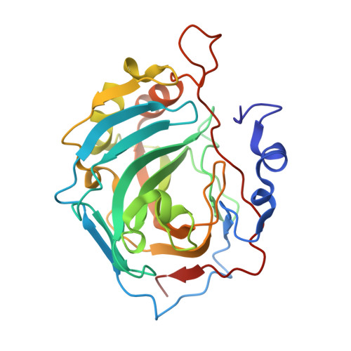
Protein-aromatic foldamer complex crystal structure


Experimental Data Snapshot

  • Method: X-RAY DIFFRACTION
  • Resolution: 2.91 Å
  • R-Value Free: 
    0.210 (Depositor), 0.217 (DCC) 
  • R-Value Work: 
    0.171 (Depositor), 0.181 (DCC) 
  • R-Value Observed: 
    0.173 (Depositor) 

Starting Model: experimental
View more details

wwPDB Validation 3D Report Full Report

Validation slider image for 6HZX

This is version 3.1 of the entry. See complete history

Literature

Structure Elucidation of Helical Aromatic Foldamer-Protein Complexes with Large Contact Surface Areas.

Reddy, P.S.Langlois d'Estaintot, B.Granier, T.Mackereth, C.D.Fischer, L.Huc, I.

(2019) Chemistry 25: 11042-11047

  • DOI: https://doi.org/10.1002/chem.201902942
  • Primary Citation Related Structures: 
    6HZX, 6Q9T

  • PubMed Abstract: 

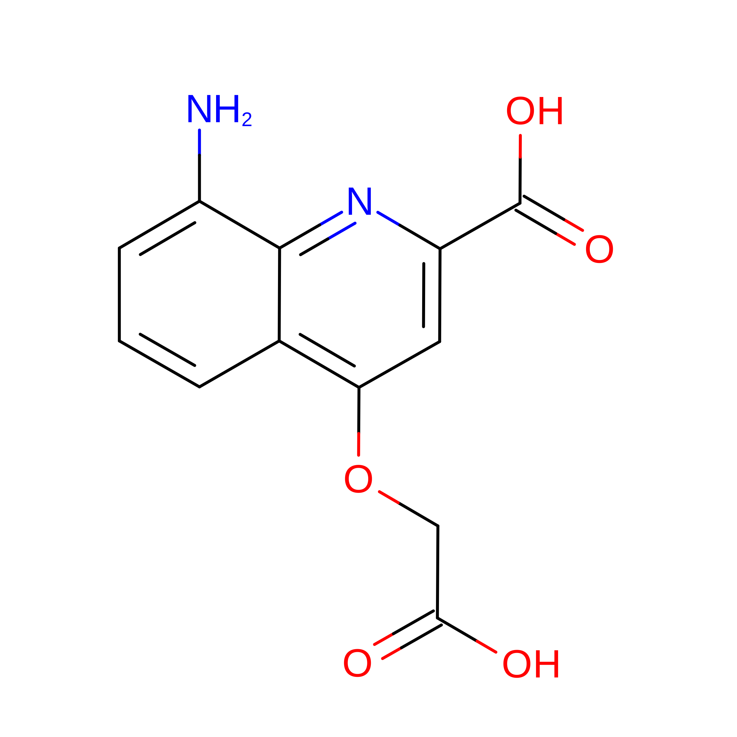
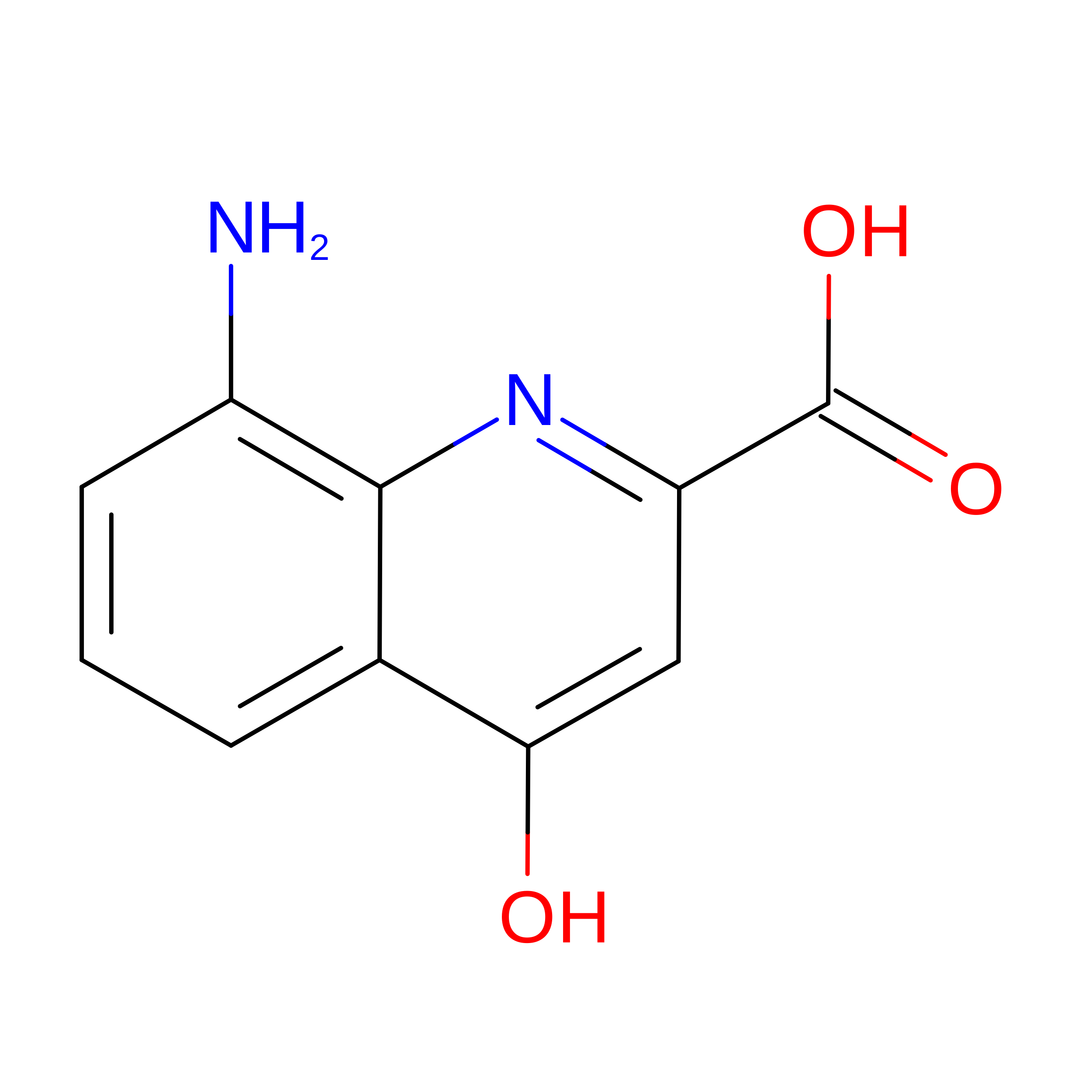
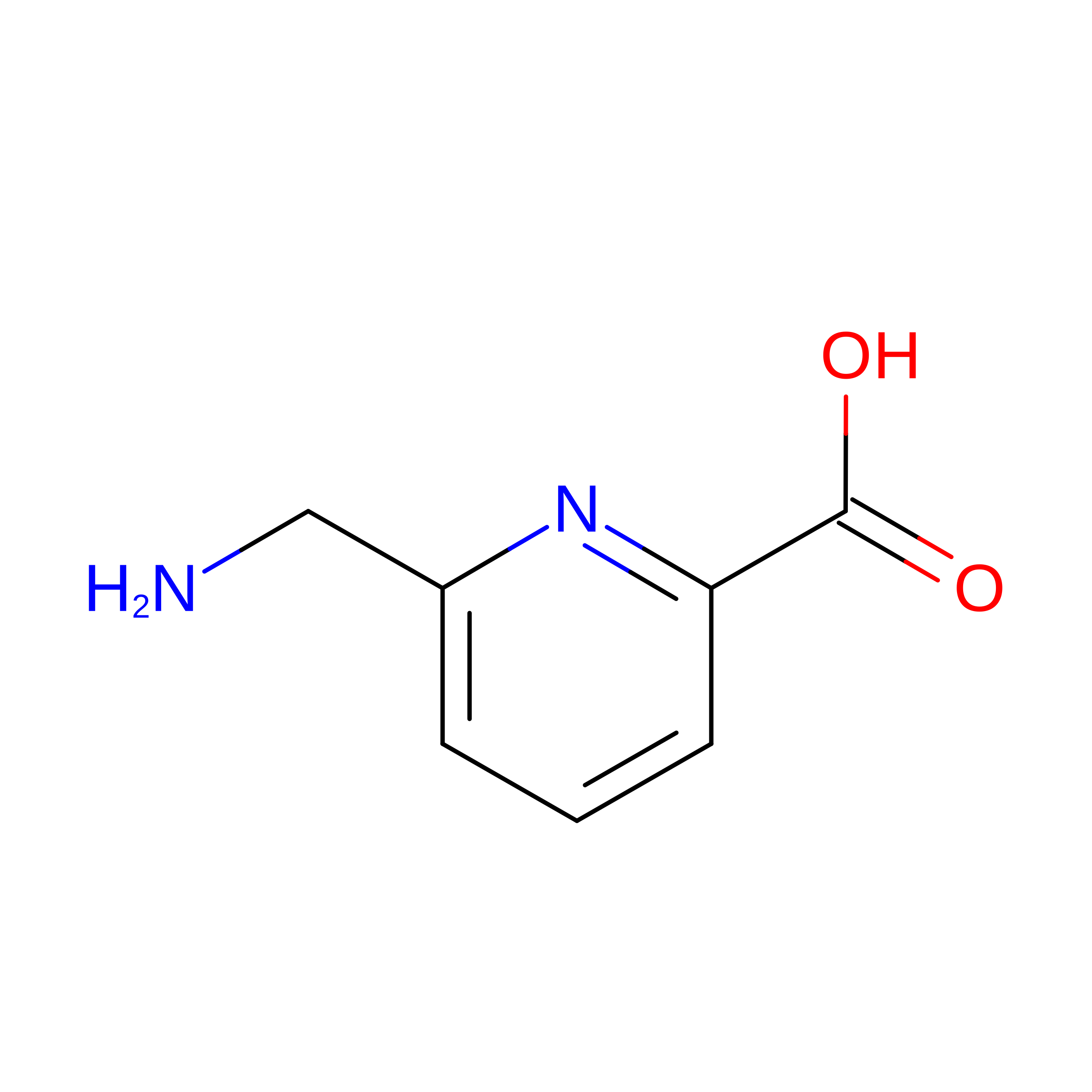
    The development of large synthetic ligands could be useful to target the sizeable surface areas involved in protein-protein interactions. Herein, we present long helical aromatic oligoamide foldamers bearing proteinogenic side chains that cover up to 450 Å 2 of the human carbonic anhydrase II (HCA) surface. The foldamers are composed of aminoquinolinecarboxylic acids bearing proteinogenic side chains and of more flexible aminomethyl-pyridinecarboxylic acids that enhance helix handedness dynamics. Crystal structures of HCA-foldamer complexes were obtained with a 9- and a 14-mer both showing extensive protein-foldamer hydrophobic contacts. In addition, foldamer-foldamer interactions seem to be prevalent in the crystal packing, leading to the peculiar formation of an HCA superhelix wound around a rod of stacked foldamers. Solution studies confirm the positioning of the foldamer at the protein surface as well as a dimerization of the complexes.


  • Organizational Affiliation
    • CBMN (UMR5248), Univ. Bordeaux-CNRS-INP, Institut Européen de Chimie et Biologie, 2 rue Escarpit, 33600, Pessac, France.

Macromolecule Content 

  • Total Structure Weight: 67.22 kDa 
  • Atom Count: 4,687 
  • Modeled Residue Count: 558 
  • Deposited Residue Count: 558 
  • Unique protein chains: 2

Macromolecules

Find similar proteins by:|  3D Structure
Entity ID: 1
MoleculeChains  Sequence LengthOrganismDetailsImage
Carbonic anhydrase 2
A, B
257Homo sapiensMutation(s): 0 
Gene Names: CA2
EC: 4.2.1.1 (PDB Primary Data), 4.2.1.69 (UniProt)
UniProt & NIH Common Fund Data Resources
Find proteins for P00918 (Homo sapiens)
Explore P00918 
Go to UniProtKB:  P00918
PHAROS:  P00918
GTEx:  ENSG00000104267 
Entity Groups
Sequence Clusters30% Identity50% Identity70% Identity90% Identity95% Identity100% Identity
UniProt GroupP00918
Sequence Annotations
Expand
Reference Sequence
Find similar proteins by:  Sequence   |   3D Structure  
Entity ID: 2
MoleculeChains  Sequence LengthOrganismDetailsImage
Aromatic foldamer
C, D, E, F
11synthetic constructMutation(s): 0 
Sequence Annotations
Expand
Reference Sequence

Small Molecules

Modified Residues  6 Unique
IDChains TypeFormula2D DiagramParent
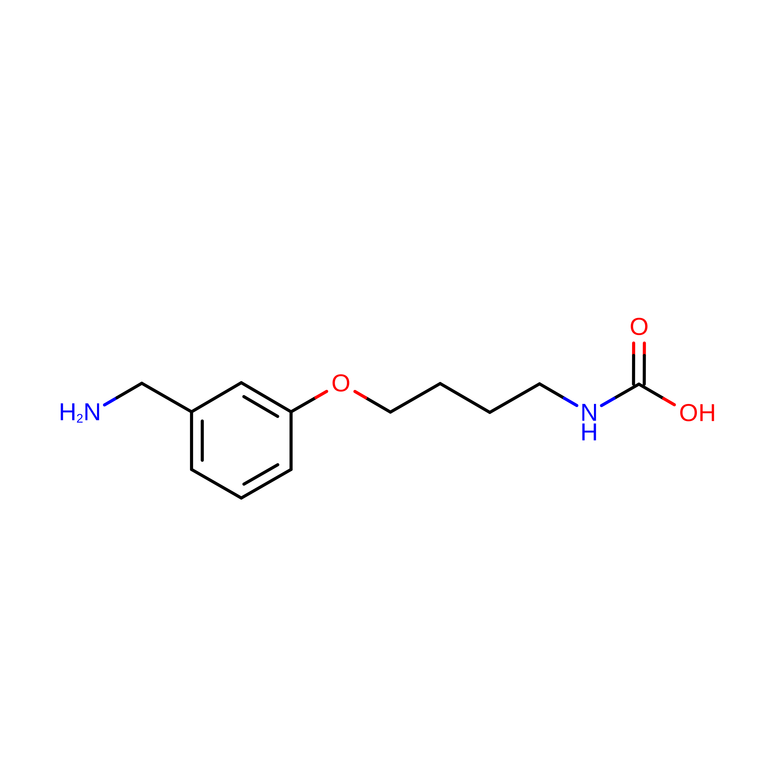
A1IJ4
Query on A1IJ4
C, D, E, F
L-PEPTIDE LINKINGC12 H18 N2 O3

--

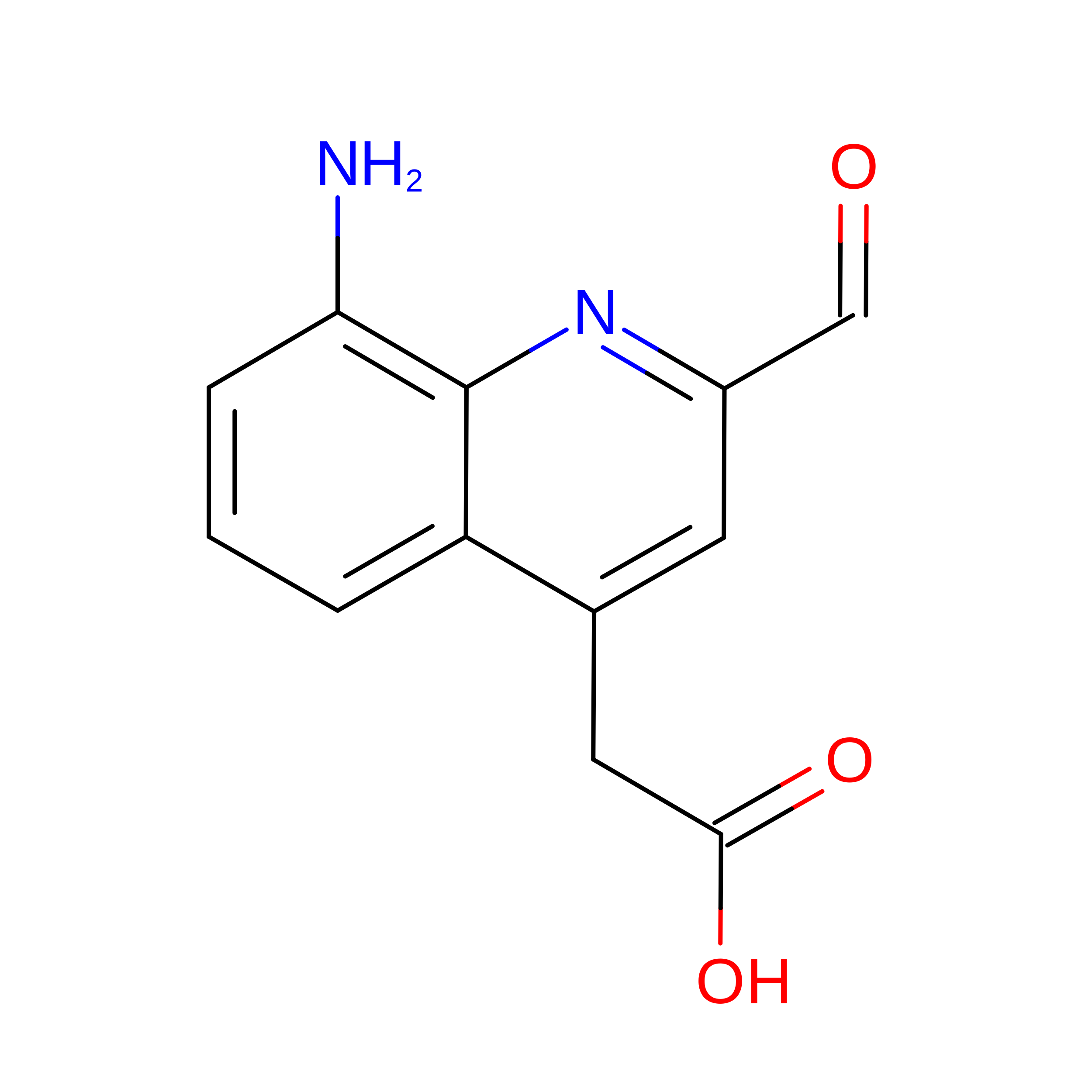
QDD
Query on QDD
C, D, E, F
L-PEPTIDE LINKINGC12 H10 N2 O4

--

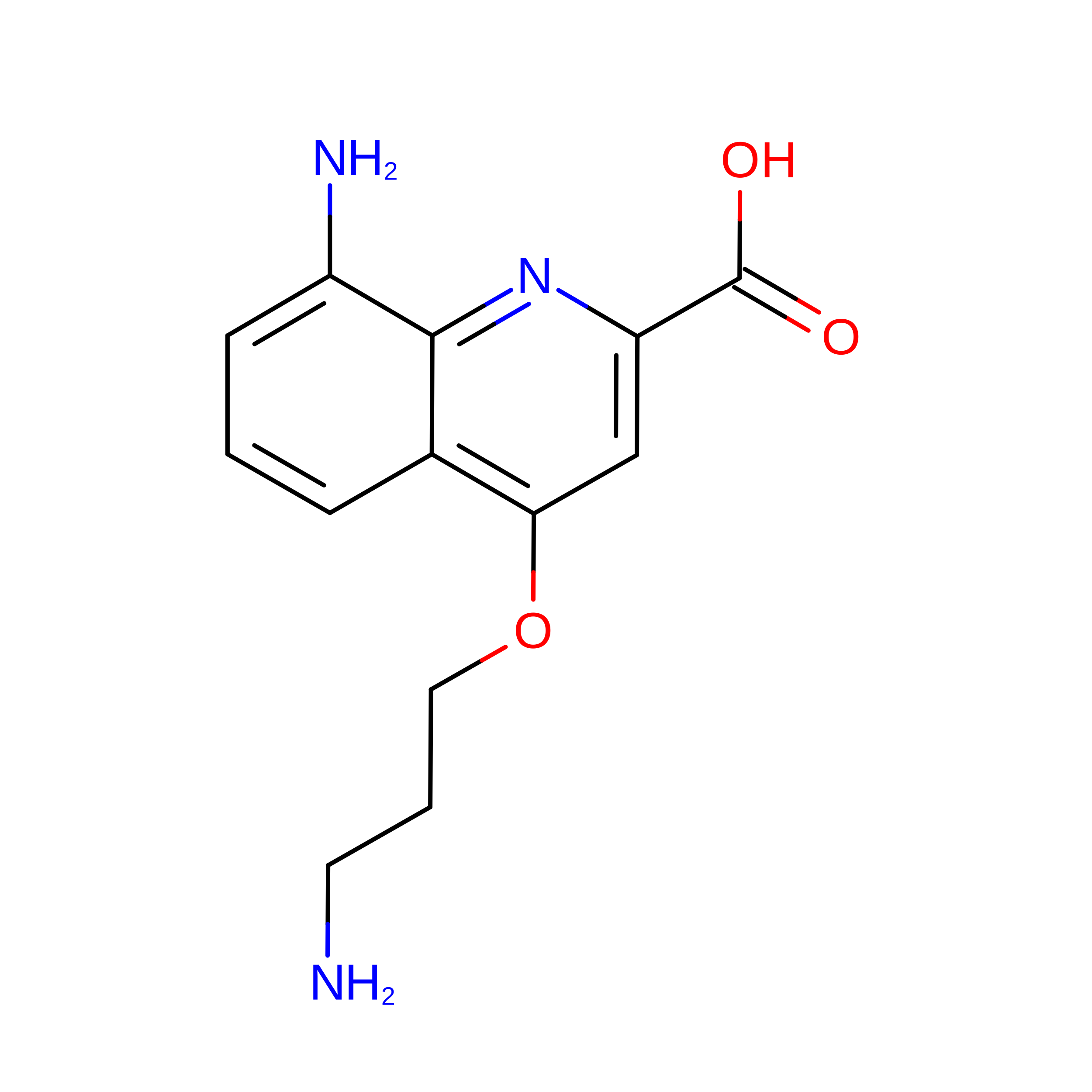
QUK
Query on QUK
C, D, E, F
L-PEPTIDE LINKINGC13 H15 N3 O3

--

QVE
Query on QVE
C, D, E, F
L-PEPTIDE LINKINGC12 H10 N2 O5

--

QVS
Query on QVS
C, D, E, F
L-PEPTIDE LINKINGC10 H8 N2 O3

--

ZY9
Query on ZY9
C, D, E, F
L-PEPTIDE LINKINGC7 H8 N2 O2

--

Experimental Data & Validation

Experimental Data

  • Method: X-RAY DIFFRACTION
  • Resolution: 2.91 Å
  • R-Value Free:  0.210 (Depositor), 0.217 (DCC) 
  • R-Value Work:  0.171 (Depositor), 0.181 (DCC) 
  • R-Value Observed: 0.173 (Depositor) 
Space Group: P 43
Unit Cell:
Length ( Å )Angle ( ˚ )
a = 82.64α = 90
b = 82.64β = 90
c = 106.39γ = 90
Software Package:
Software NamePurpose
REFMACrefinement
XDSdata reduction
XSCALEdata scaling
PHASERphasing
PDB_EXTRACTdata extraction

Structure Validation

View Full Validation Report


Entry History 

Deposition Data

Revision History  (Full details and data files)

  • Version 1.0: 2019-09-18
    Type: Initial release
  • Version 2.0: 2023-11-15
    Changes: Atomic model, Data collection, Database references, Derived calculations, Refinement description
  • Version 2.1: 2024-01-24
    Changes: Refinement description
  • Version 3.0: 2024-08-14
    Changes: Advisory, Atomic model, Data collection, Database references, Derived calculations, Non-polymer description, Other, Polymer sequence, Refinement description, Source and taxonomy, Structure summary
  • Version 3.1: 2024-11-06
    Changes: Derived calculations, Structure summary