6I0L | pdb_00006i0l

Crystal structure of human carbonic anhydrase I in complex with the 1-[4-chloro-3-(trifluoromethyl)phenyl]-3-[2-(4-sulfamoylphenyl)ethyl]urea inhibitor


Experimental Data Snapshot

  • Method: X-RAY DIFFRACTION
  • Resolution: 1.40 Å
  • R-Value Free: 
    0.237 (Depositor), 0.237 (DCC) 
  • R-Value Work: 
    0.204 (Depositor), 0.204 (DCC) 
  • R-Value Observed: 
    0.206 (Depositor) 

Starting Model: experimental
View more details

wwPDB Validation 3D Report Full Report

Validation slider image for 6I0L

Ligand Structure Quality Assessment 


This is version 1.2 of the entry. See complete history

Literature

Carbonic anhydrase inhibitors based on sorafenib scaffold: Design, synthesis, crystallographic investigation and effects on primary breast cancer cells.

Bozdag, M.Ferraroni, M.Ward, C.Carta, F.Bua, S.Angeli, A.Langdon, S.P.Kunkler, I.H.Al-Tamimi, A.S.Supuran, C.T.

(2019) Eur J Med Chem 182: 111600-111600

  • DOI: https://doi.org/10.1016/j.ejmech.2019.111600
  • Primary Citation Related Structures: 
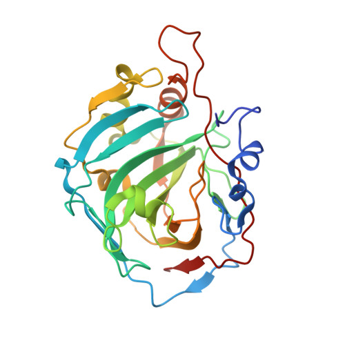
    6I0J, 6I0L

  • PubMed Abstract: 

    Carbonic anhydrase inhibitors (CAIs) of the sulfonamide, sulfamate and coumarin classes bearing the phenylureido tail found in the clinically used drug Sorafenib, a multikinase inhibitor actually used for the management of hepatocellular carcinomas, are reported. All compounds were assayed on human (h) CA isoforms I, II, VII and IX, involved in various pathologies. Among the sulfonamides, several compounds were selective for inhibiting hCA IX, with K I values in the low nanomolar ranges (i.e. 0.7-30.2 nM). We explored the binding modes of such compounds by means of X-ray crystallographic studies on isoform hCA I in adduct with one sulfonamide and a sulfamate inhibitor. Antiproliferative properties of some sulfamates on breast tumor cell lines were also investigated.


  • Organizational Affiliation
    • University of Florence, NEUROFARBA Dept, Sezione di Scienze Farmaceutiche e Nutraceutiche, Via Ugo Schiff 6, 50019, Sesto Fiorentino, Florence, Italy. Electronic address: bozdag.murat@unifi.it.

Macromolecule Content 

  • Total Structure Weight: 59.12 kDa 
  • Atom Count: 4,618 
  • Modeled Residue Count: 514 
  • Deposited Residue Count: 522 
  • Unique protein chains: 1

Macromolecules

Find similar proteins by:|  3D Structure
Entity ID: 1
MoleculeChains  Sequence LengthOrganismDetailsImage
Carbonic anhydrase 1
A, B
261Homo sapiensMutation(s): 0 
EC: 4.2.1.1 (PDB Primary Data), 4.2.1.69 (UniProt)
UniProt & NIH Common Fund Data Resources
Find proteins for P00915 (Homo sapiens)
Explore P00915 
Go to UniProtKB:  P00915
PHAROS:  P00915
GTEx:  ENSG00000133742 
Entity Groups
Sequence Clusters30% Identity50% Identity70% Identity90% Identity95% Identity100% Identity
UniProt GroupP00915
Sequence Annotations
Expand
Reference Sequence

Small Molecules

Ligands 4 Unique
IDChains Name / Formula / InChI Key2D Diagram3D Interactions
GZH
(Subject of Investigation/LOI)

Query on GZH


Download:Ideal Coordinates CCD File
D [auth A],
H [auth B]
1-[4-chloranyl-3-(trifluoromethyl)phenyl]-3-[2-(4-sulfamoylphenyl)ethyl]urea
C16 H15 Cl F3 N3 O3 S
FBVAPDWPSNARKD-UHFFFAOYSA-N
GOL

Query on GOL


Download:Ideal Coordinates CCD File
E [auth A],
I [auth B],
J [auth B]
GLYCEROL
C3 H8 O3
PEDCQBHIVMGVHV-UHFFFAOYSA-N
ZN

Query on ZN


Download:Ideal Coordinates CCD File
C [auth A],
G [auth B]
ZINC ION
Zn
PTFCDOFLOPIGGS-UHFFFAOYSA-N
ACT

Query on ACT


Download:Ideal Coordinates CCD File
F [auth A]ACETATE ION
C2 H3 O2
QTBSBXVTEAMEQO-UHFFFAOYSA-M
Binding Affinity Annotations 
IDSourceBinding Affinity
GZH BindingDB:  6I0L Ki: 913 (nM) from 1 assay(s)
ACT BindingDB:  6I0L Ki: 1.08e+4 (nM) from 1 assay(s)

Experimental Data & Validation

Experimental Data

  • Method: X-RAY DIFFRACTION
  • Resolution: 1.40 Å
  • R-Value Free:  0.237 (Depositor), 0.237 (DCC) 
  • R-Value Work:  0.204 (Depositor), 0.204 (DCC) 
  • R-Value Observed: 0.206 (Depositor) 
Space Group: P 21 21 21
Unit Cell:
Length ( Å )Angle ( ˚ )
a = 62.461α = 90
b = 70.453β = 90
c = 121.56γ = 90
Software Package:
Software NamePurpose
XSCALEdata scaling
REFMACrefinement
PDB_EXTRACTdata extraction
XDSdata reduction
REFMACphasing

Structure Validation

View Full Validation Report


Ligand Structure Quality Assessment 


Entry History 

Deposition Data

Revision History  (Full details and data files)

  • Version 1.0: 2019-11-20
    Type: Initial release
  • Version 1.1: 2020-12-02
    Changes: Database references, Derived calculations
  • Version 1.2: 2024-01-24
    Changes: Data collection, Database references, Refinement description