6I3V

x-ray structure of the human mitochondrial PRELID1 in complex with TRIAP1


Experimental Data Snapshot

  • Method: X-RAY DIFFRACTION
  • Resolution: 1.98 Å
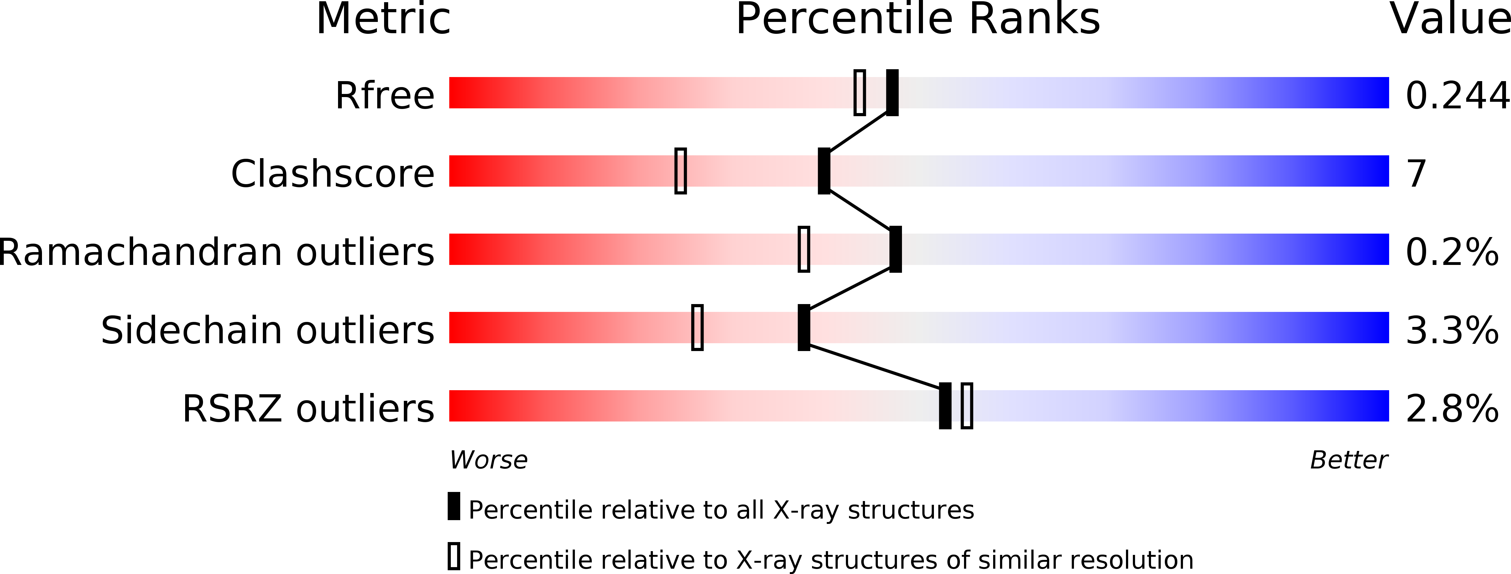
  • R-Value Free: 0.245 
  • R-Value Work: 0.205 
  • R-Value Observed: 0.207 

wwPDB Validation   3D Report Full Report


Ligand Structure Quality Assessment 


This is version 1.1 of the entry. See complete history


Literature

Structural determinants of lipid specificity within Ups/PRELI lipid transfer proteins.

Miliara, X.Tatsuta, T.Berry, J.L.Rouse, S.L.Solak, K.Chorev, D.S.Wu, D.Robinson, C.V.Matthews, S.Langer, T.

(2019) Nat Commun 10: 1130-1130

  • DOI: https://doi.org/10.1038/s41467-019-09089-x
  • Primary Citation of Related Structures:  
    6I3V, 6I3Y, 6I4Y

  • PubMed Abstract: 

    Conserved lipid transfer proteins of the Ups/PRELI family regulate lipid accumulation in mitochondria by shuttling phospholipids in a lipid-specific manner across the intermembrane space. Here, we combine structural analysis, unbiased genetic approaches in yeast and molecular dynamics simulations to unravel determinants of lipid specificity within the conserved Ups/PRELI family. We present structures of human PRELID1-TRIAP1 and PRELID3b-TRIAP1 complexes, which exert lipid transfer activity for phosphatidic acid and phosphatidylserine, respectively. Reverse yeast genetic screens identify critical amino acid exchanges that broaden and swap their lipid specificities. We find that amino acids involved in head group recognition and the hydrophobicity of flexible loops regulate lipid entry into the binding cavity. Molecular dynamics simulations reveal different membrane orientations of PRELID1 and PRELID3b during the stepwise release of lipids. Our experiments thus define the structural determinants of lipid specificity and the dynamics of lipid interactions by Ups/PRELI proteins.


  • Organizational Affiliation

    Department of Life Sciences, Imperial College London, Sir Ernst Chain Building, South Kensington, London, SW7 2AZ, UK.


Macromolecules
Find similar proteins by:  (by identity cutoff)  |  3D Structure
Entity ID: 1
MoleculeChains Sequence LengthOrganismDetailsImage
PRELI domain-containing protein 1, mitochondrialA [auth F],
B
182Homo sapiensMutation(s): 0 
Gene Names: PRELID1PRELICGI-106SBBI12
UniProt & NIH Common Fund Data Resources
Find proteins for Q9Y255 (Homo sapiens)
Explore Q9Y255 
Go to UniProtKB:  Q9Y255
PHAROS:  Q9Y255
GTEx:  ENSG00000169230 
Entity Groups  
Sequence Clusters30% Identity50% Identity70% Identity90% Identity95% Identity100% Identity
UniProt GroupQ9Y255
Sequence Annotations
Expand
  • Reference Sequence
Find similar proteins by:  (by identity cutoff)  |  3D Structure
Entity ID: 2
MoleculeChains Sequence LengthOrganismDetailsImage
TP53-regulated inhibitor of apoptosis 1C [auth A],
D [auth C]
69Homo sapiensMutation(s): 0 
Gene Names: TRIAP115E1.1HSPC132
UniProt & NIH Common Fund Data Resources
Find proteins for O43715 (Homo sapiens)
Explore O43715 
Go to UniProtKB:  O43715
PHAROS:  O43715
GTEx:  ENSG00000170855 
Entity Groups  
Sequence Clusters30% Identity50% Identity70% Identity90% Identity95% Identity100% Identity
UniProt GroupO43715
Sequence Annotations
Expand
  • Reference Sequence
Small Molecules
Ligands 3 Unique
IDChains Name / Formula / InChI Key2D Diagram3D Interactions
MYR
Query on MYR

Download Ideal Coordinates CCD File 
E [auth F],
I [auth B]
MYRISTIC ACID
C14 H28 O2
TUNFSRHWOTWDNC-UHFFFAOYSA-N
CL
Query on CL

Download Ideal Coordinates CCD File 
F
G [auth F]
H [auth F]
M [auth B]
N [auth B]
F,
G [auth F],
H [auth F],
M [auth B],
N [auth B],
O [auth B],
P [auth B],
Q [auth B],
R [auth B],
S [auth B],
T [auth B],
U [auth B],
V [auth C]
CHLORIDE ION
Cl
VEXZGXHMUGYJMC-UHFFFAOYSA-M
NA
Query on NA

Download Ideal Coordinates CCD File 
J [auth B],
K [auth B],
L [auth B]
SODIUM ION
Na
FKNQFGJONOIPTF-UHFFFAOYSA-N
Experimental Data & Validation

Experimental Data

  • Method: X-RAY DIFFRACTION
  • Resolution: 1.98 Å
  • R-Value Free: 0.245 
  • R-Value Work: 0.205 
  • R-Value Observed: 0.207 
  • Space Group: P 63 2 2
Unit Cell:
Length ( Å )Angle ( ˚ )
a = 127.312α = 90
b = 127.312β = 90
c = 177.39γ = 120
Software Package:
Software NamePurpose
PHENIXrefinement
PDB_EXTRACTdata extraction
XDSdata reduction
SCALAdata scaling
PHASERphasing

Structure Validation

View Full Validation Report


Ligand Structure Quality Assessment 


Entry History & Funding Information

Deposition Data


Funding OrganizationLocationGrant Number
United Kingdom--

Revision History  (Full details and data files)

  • Version 1.0: 2019-03-20
    Type: Initial release
  • Version 1.1: 2024-11-20
    Changes: Data collection, Database references, Structure summary