Discovery and Optimization of wt-RET/KDR-Selective Inhibitors of RETV804MKinase.
Newton, R., Waszkowycz, B., Seewooruthun, C., Burschowsky, D., Richards, M., Hitchin, S., Begum, H., Watson, A., French, E., Hamilton, N., Jones, S., Lin, L.Y., Waddell, I., Echalier, A., Bayliss, R., Jordan, A.M., Ogilvie, D.(2020) ACS Med Chem Lett 11: 497-505
- PubMed: 32292556 Search on PubMedSearch on PubMed Central
- DOI: https://doi.org/10.1021/acsmedchemlett.9b00615
- Primary Citation Related Structures:
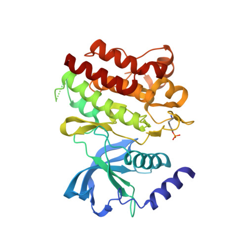
6I82, 6I83 - PubMed Abstract:
A combination of focused library and virtual screening, hit expansion, and rational design has resulted in the development of a series of inhibitors of RET V804M kinase, the anticipated drug-resistant mutant of RET kinase. These agents do not inhibit the wild type (wt) isoforms of RET or KDR and therefore offer a potential adjunct to RET inhibitors currently undergoing clinical evaluation.
- Drug Discovery Unit, Cancer Research UK, Manchester Institute, University of Manchester, Alderley Park, Macclesfield SK10 4TG, U.K.
Organizational Affiliation: