6IS0

Crystal structure of the zebrafish cap-specific adenosine methyltransferase bound to SAH and m7G-capped RNA


Experimental Data Snapshot

  • Method: X-RAY DIFFRACTION
  • Resolution: 1.80 Å
  • R-Value Free: 
    0.214 (Depositor), 0.220 (DCC) 
  • R-Value Work: 
    0.177 (Depositor), 0.180 (DCC) 
  • R-Value Observed: 
    0.179 (Depositor) 

Starting Model: experimental
View more details

wwPDB Validation   3D Report Full Report


Ligand Structure Quality Assessment 

Created with Raphaël 2.3.0Worse 01 BetterLigand structure goodness of fit to experimental dataBest fitted M7GClick on this verticalbar to view detailsBest fitted SAHClick on this verticalbar to view details

This is version 2.0 of the entry. See complete history


Literature

Cap-specific terminal N 6 -methylation of RNA by an RNA polymerase II-associated methyltransferase.

Akichika, S.Hirano, S.Shichino, Y.Suzuki, T.Nishimasu, H.Ishitani, R.Sugita, A.Hirose, Y.Iwasaki, S.Nureki, O.Suzuki, T.

(2019) Science 363

  • DOI: https://doi.org/10.1126/science.aav0080
  • Primary Citation of Related Structures:  
    6IRV, 6IRW, 6IRX, 6IRY, 6IRZ, 6IS0

  • PubMed Abstract: 

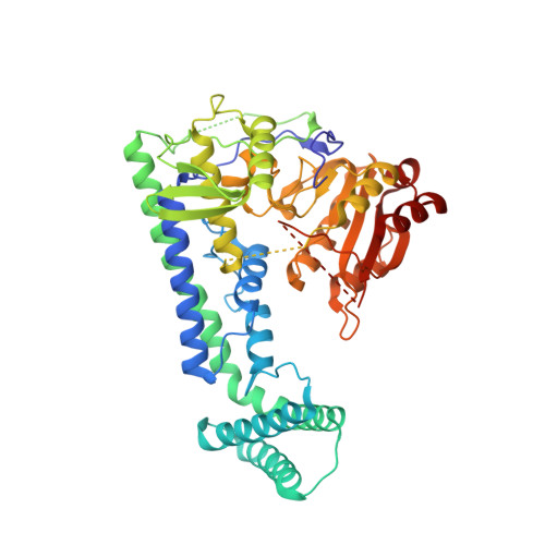
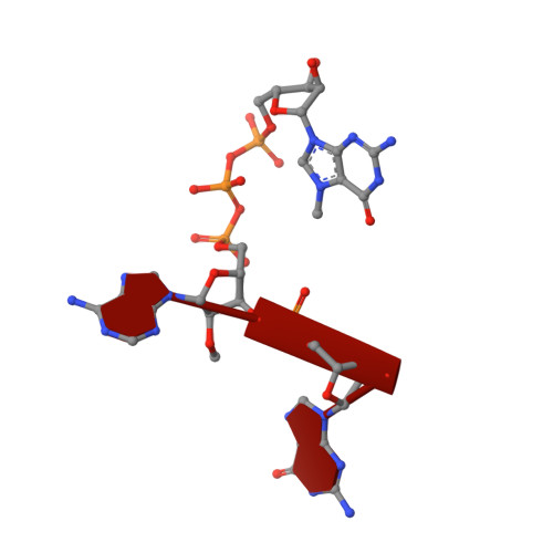
    N 6 -methyladenosine (m 6 A), a major modification of messenger RNAs (mRNAs), plays critical roles in RNA metabolism and function. In addition to the internal m 6 A, N 6 , 2'- O -dimethyladenosine (m 6 Am) is present at the transcription start nucleotide of capped mRNAs in vertebrates. However, its biogenesis and functional role remain elusive. Using a reverse genetics approach, we identified PCIF1, a factor that interacts with the serine-5-phosphorylated carboxyl-terminal domain of RNA polymerase II, as a cap-specific adenosine methyltransferase (CAPAM) responsible for N 6 -methylation of m 6 Am. The crystal structure of CAPAM in complex with substrates revealed the molecular basis of cap-specific m 6 A formation. A transcriptome-wide analysis revealed that N 6 -methylation of m 6 Am promotes the translation of capped mRNAs. Thus, a cap-specific m 6 A writer promotes translation of mRNAs starting from m 6 Am.


  • Organizational Affiliation

    Department of Chemistry and Biotechnology, Graduate School of Engineering, The University of Tokyo, 7-3-1 Hongo, Bunkyo-ku, Tokyo 113-8656, Japan.


Macromolecules

Find similar proteins by:  (by identity cutoff)  |  3D Structure
Entity ID: 1
MoleculeChains Sequence LengthOrganismDetailsImage
PDX1 C-terminal-inhibiting factor 1496Danio rerioMutation(s): 2 
Gene Names: pcif1
EC: 2.1.1.62
UniProt
Find proteins for A0A0R4IKJ1 (Danio rerio)
Explore A0A0R4IKJ1 
Go to UniProtKB:  A0A0R4IKJ1
Entity Groups  
Sequence Clusters30% Identity50% Identity70% Identity90% Identity95% Identity100% Identity
UniProt GroupA0A0R4IKJ1
Sequence Annotations
Expand
  • Reference Sequence
Small Molecules
Ligands 4 Unique
IDChains Name / Formula / InChI Key2D Diagram3D Interactions
M7G
Query on M7G

Download Ideal Coordinates CCD File 
D [auth A]7N-METHYL-8-HYDROGUANOSINE-5'-DIPHOSPHATE
C11 H18 N5 O11 P2
SBASPRRECYVBRF-KQYNXXCUSA-O
SAH
Query on SAH

Download Ideal Coordinates CCD File 
C [auth A]S-ADENOSYL-L-HOMOCYSTEINE
C14 H20 N6 O5 S
ZJUKTBDSGOFHSH-WFMPWKQPSA-N
B3P
Query on B3P

Download Ideal Coordinates CCD File 
I [auth A]2-[3-(2-HYDROXY-1,1-DIHYDROXYMETHYL-ETHYLAMINO)-PROPYLAMINO]-2-HYDROXYMETHYL-PROPANE-1,3-DIOL
C11 H26 N2 O6
HHKZCCWKTZRCCL-UHFFFAOYSA-N
EDO
Query on EDO

Download Ideal Coordinates CCD File 
E [auth A],
F [auth A],
G [auth A],
H [auth A]
1,2-ETHANEDIOL
C2 H6 O2
LYCAIKOWRPUZTN-UHFFFAOYSA-N
Experimental Data & Validation

Experimental Data

  • Method: X-RAY DIFFRACTION
  • Resolution: 1.80 Å
  • R-Value Free:  0.214 (Depositor), 0.220 (DCC) 
  • R-Value Work:  0.177 (Depositor), 0.180 (DCC) 
  • R-Value Observed: 0.179 (Depositor) 
Space Group: P 21 21 21
Unit Cell:
Length ( Å )Angle ( ˚ )
a = 74.219α = 90
b = 84.035β = 90
c = 92.97γ = 90
Software Package:
Software NamePurpose
Aimlessdata scaling
PHENIXrefinement
PDB_EXTRACTdata extraction
XDSdata reduction
MOLREPphasing

Structure Validation

View Full Validation Report


Ligand Structure Quality Assessment 

Created with Raphaël 2.3.0Worse 01 BetterLigand structure goodness of fit to experimental dataBest fitted M7GClick on this verticalbar to view detailsBest fitted SAHClick on this verticalbar to view details

Entry History 

Deposition Data

Revision History  (Full details and data files)

  • Version 1.0: 2018-12-05
    Type: Initial release
  • Version 1.1: 2019-01-23
    Changes: Data collection, Database references
  • Version 1.2: 2020-10-28
    Changes: Database references
  • Version 1.3: 2023-11-22
    Changes: Data collection, Database references, Refinement description
  • Version 2.0: 2024-03-06
    Changes: Data collection, Non-polymer description, Structure summary