6J17

ATPase


Experimental Data Snapshot

  • Method: X-RAY DIFFRACTION
  • Resolution: 1.98 Å
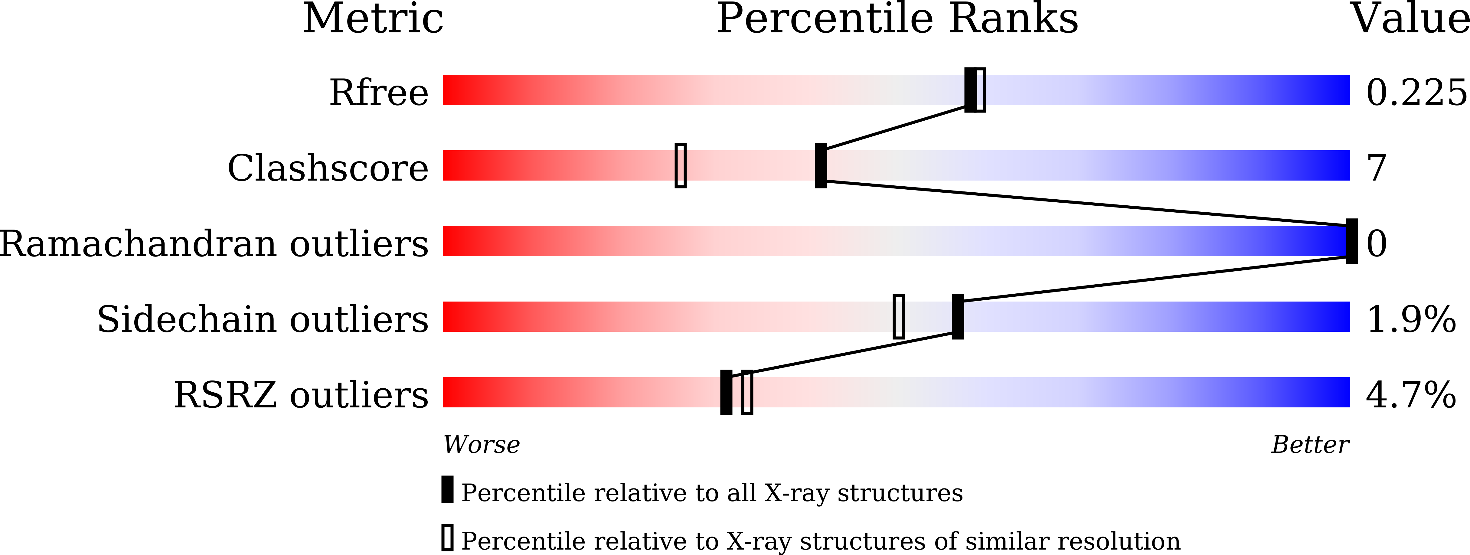
  • R-Value Free: 0.228 
  • R-Value Work: 0.181 
  • R-Value Observed: 0.183 

Starting Model: experimental
View more details

wwPDB Validation   3D Report Full Report


Ligand Structure Quality Assessment 


This is version 1.2 of the entry. See complete history


Literature

Structural insights into substrate recognition by the type VII secretion system.

Wang, S.Zhou, K.Yang, X.Zhang, B.Zhao, Y.Xiao, Y.Yang, X.Yang, H.Guddat, L.W.Li, J.Rao, Z.

(2020) Protein Cell 11: 124-137

  • DOI: https://doi.org/10.1007/s13238-019-00671-z
  • Primary Citation of Related Structures:  
    6J17, 6J18, 6J19, 6JD4, 6JD5

  • PubMed Abstract: 

    Type VII secretion systems (T7SSs) are found in many disease related bacteria including Mycobacterium tuberculosis (Mtb). ESX-1 [early secreted antigen 6 kilodaltons (ESAT-6) system 1] is one of the five subtypes (ESX-1~5) of T7SSs in Mtb, where it delivers virulence factors into host macrophages during infection. However, little is known about the molecular details as to how this occurs. Here, we provide high-resolution crystal structures of the C-terminal ATPase 3 domains of EccC subunits from four different Mtb T7SS subtypes. These structures adopt a classic RecA-like ɑ/β fold with a conserved Mg-ATP binding site. The structure of EccCb1 in complex with the C-terminal peptide of EsxB identifies the location of substrate recognition site and shows how the specific signaling module "LxxxMxF" for Mtb ESX-1 binds to this site resulting in a translation of the bulge loop. A comparison of all the ATPase 3 structures shows there are significant differences in the shape and composition of the signal recognition pockets across the family, suggesting that distinct signaling sequences of substrates are required to be specifically recognized by different T7SSs. A hexameric model of the EccC-ATPase 3 is proposed and shows the recognition pocket is located near the central substrate translocation channel. The diameter of the channel is ~25-Å, with a size that would allow helix-bundle shaped substrate proteins to bind and pass through. Thus, our work provides new molecular insights into substrate recognition for Mtb T7SS subtypes and also a possible transportation mechanism for substrate and/or virulence factor secretion.


  • Organizational Affiliation

    Shanghai Institute for Advanced Immunochemical Studies and School of Life Science and Technology, ShanghaiTech University, Shanghai, 201210, China.


Macromolecules
Find similar proteins by:  (by identity cutoff)  |  3D Structure
Entity ID: 1
MoleculeChains Sequence LengthOrganismDetailsImage
ESX-3 secretion system protein EccC3324Mycobacterium tuberculosis H37RvMutation(s): 0 
Gene Names: eccC3Rv0284
UniProt
Find proteins for P9WNA9 (Mycobacterium tuberculosis (strain ATCC 25618 / H37Rv))
Explore P9WNA9 
Go to UniProtKB:  P9WNA9
Entity Groups  
Sequence Clusters30% Identity50% Identity70% Identity90% Identity95% Identity100% Identity
UniProt GroupP9WNA9
Sequence Annotations
Expand
  • Reference Sequence
Experimental Data & Validation

Experimental Data

  • Method: X-RAY DIFFRACTION
  • Resolution: 1.98 Å
  • R-Value Free: 0.228 
  • R-Value Work: 0.181 
  • R-Value Observed: 0.183 
  • Space Group: P 21 21 21
Unit Cell:
Length ( Å )Angle ( ˚ )
a = 53.363α = 90
b = 56.628β = 90
c = 93.532γ = 90
Software Package:
Software NamePurpose
PHENIXrefinement
SCALEPACKdata scaling
PDB_EXTRACTdata extraction
HKL-2000data reduction
PHASERphasing

Structure Validation

View Full Validation Report


Ligand Structure Quality Assessment 


Entry History & Funding Information

Deposition Data


Funding OrganizationLocationGrant Number
Ministry of Science and Technology (China)China2017YFC0840300
Chinese Academy of SciencesChinaXDB08020200
Ministry of Science and Technology (China)China2014CB542800
Ministry of Science and Technology (China)China2014CBA02003
National Natural Science Foundation of ChinaChina813300237
National Natural Science Foundation of ChinaChina31500607
National Natural Science Foundation of ChinaChina81520108019

Revision History  (Full details and data files)

  • Version 1.0: 2019-12-04
    Type: Initial release
  • Version 1.1: 2020-01-29
    Changes: Database references
  • Version 1.2: 2023-11-22
    Changes: Data collection, Database references, Derived calculations, Refinement description