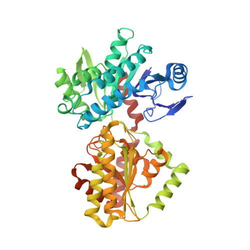
Crystal structures of Magnaporthe oryzae trehalose-6-phosphate synthase (MoTps1) suggest a model for catalytic process of Tps1.
Wang, S., Zhao, Y., Yi, L., Shen, M., Wang, C., Zhang, X., Yang, J., Peng, Y.L., Wang, D., Liu, J.(2019) Biochem J 476: 3227-3240
- PubMed: 31455720
- DOI: https://doi.org/10.1042/BCJ20190289
- Primary Citation of Related Structures:
6JAK, 6JBI, 6JBR, 6JBW - PubMed Abstract:
Trehalose-6-phosphate (T6P) synthase (Tps1) catalyzes the formation of T6P from UDP-glucose (UDPG) (or GDPG, etc.) and glucose-6-phosphate (G6P), and structural basis of this process has not been well studied. MoTps1 (Magnaporthe oryzae Tps1) plays a critical role in carbon and nitrogen metabolism, but its structural information is unknown. Here we present the crystal structures of MoTps1 apo, binary (with UDPG) and ternary (with UDPG/G6P or UDP/T6P) complexes. MoTps1 consists of two modified Rossmann-fold domains and a catalytic center in-between. Unlike Escherichia coli OtsA (EcOtsA, the Tps1 of E. coli), MoTps1 exists as a mixture of monomer, dimer, and oligomer in solution. Inter-chain salt bridges, which are not fully conserved in EcOtsA, play primary roles in MoTps1 oligomerization. Binding of UDPG by MoTps1 C-terminal domain modifies the substrate pocket of MoTps1. In the MoTps1 ternary complex structure, UDP and T6P, the products of UDPG and G6P, are detected, and substantial conformational rearrangements of N-terminal domain, including structural reshuffling (β3-β4 loop to α0 helix) and movement of a 'shift region' towards the catalytic centre, are observed. These conformational changes render MoTps1 to a 'closed' state compared with its 'open' state in apo or UDPG complex structures. By solving the EcOtsA apo structure, we confirmed that similar ligand binding induced conformational changes also exist in EcOtsA, although no structural reshuffling involved. Based on our research and previous studies, we present a model for the catalytic process of Tps1. Our research provides novel information on MoTps1, Tps1 family, and structure-based antifungal drug design.
- MOA Key Laboratory of Plant Pathology, Joint International Research Laboratory of Crop Molecular Breeding, College of Plant Protection, China Agricultural University, Beijing 100193, China.
Organizational Affiliation:


