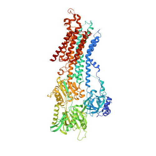
6JJU | pdb_00006jju

Structure of Ca2+ ATPase


Experimental Data Snapshot

  • Method: X-RAY DIFFRACTION
  • Resolution: 3.20 Å
  • R-Value Free: 
    0.256 (Depositor), 0.260 (DCC) 
  • R-Value Work: 
    0.211 (Depositor), 0.210 (DCC) 
  • R-Value Observed: 
    0.214 (Depositor) 

Starting Model: experimental
View more details

wwPDB Validation   3D Report Full Report


Ligand Structure Quality Assessment 

Created with Raphaël 2.3.0Worse 01 BetterLigand structure goodness of fit to experimental dataBest fitted ACPClick on this verticalbar to view details

This is version 1.1 of the entry. See complete history


Literature

Structural Basis of Sarco/Endoplasmic Reticulum Ca2+-ATPase 2b Regulation via Transmembrane Helix Interplay.

Inoue, M.Sakuta, N.Watanabe, S.Zhang, Y.Yoshikaie, K.Tanaka, Y.Ushioda, R.Kato, Y.Takagi, J.Tsukazaki, T.Nagata, K.Inaba, K.

(2019) Cell Rep 27: 1221-1230.e3

  • DOI: https://doi.org/10.1016/j.celrep.2019.03.106
  • Primary Citation of Related Structures:  
    5ZTF, 6JJU

  • PubMed Abstract: 

    Sarco/endoplasmic reticulum (ER) Ca 2+ -ATPase 2b (SERCA2b) is a ubiquitously expressed membrane protein that facilitates Ca 2+ uptake from the cytosol to the ER. SERCA2b includes a characteristic 11 th transmembrane helix (TM11) followed by a luminal tail, but the structural basis of SERCA regulation by these C-terminal segments remains unclear. Here, we determined the crystal structures of SERCA2b and its C-terminal splicing variant SERCA2a, both in the E1-2Ca 2+ -adenylyl methylenediphosphonate (AMPPCP) state. Despite discrepancies with the previously reported structural model of SERCA2b, TM11 was found to be located adjacent to TM10 and to interact weakly with a part of the L8/9 loop and the N-terminal end of TM10, thereby inhibiting the SERCA2b catalytic cycle. Accordingly, mutational disruption of the interactions between TM11 and its neighboring residues caused SERCA2b to display SERCA2a-like ATPase activity. We propose that TM11 serves as a key modulator of SERCA2b activity by fine-tuning the intramolecular interactions with other transmembrane regions.


  • Organizational Affiliation

    Institute of Multidisciplinary Research for Advanced Materials, Tohoku University, Sendai 980-8577, Japan; Core Research for Evolutional Science and Technology (CREST), Japan Science and Technology Agency, Saitama, Kawaguchi, Japan.


Macromolecules
Find similar proteins by:  (by identity cutoff)  |  3D Structure
Entity ID: 1
MoleculeChains Sequence LengthOrganismDetailsImage
Sarcoplasmic/endoplasmic reticulum calcium ATPase 21,025Homo sapiensMutation(s): 0 
Gene Names: ATP2A2ATP2B
EC: 7.2.2.10
Membrane Entity: Yes 
UniProt & NIH Common Fund Data Resources
Find proteins for P16615 (Homo sapiens)
Explore P16615 
Go to UniProtKB:  P16615
PHAROS:  P16615
GTEx:  ENSG00000174437 
Entity Groups  
Sequence Clusters30% Identity50% Identity70% Identity90% Identity95% Identity100% Identity
UniProt GroupP16615
Sequence Annotations
Expand
  • Reference Sequence
Experimental Data & Validation

Experimental Data

  • Method: X-RAY DIFFRACTION
  • Resolution: 3.20 Å
  • R-Value Free:  0.256 (Depositor), 0.260 (DCC) 
  • R-Value Work:  0.211 (Depositor), 0.210 (DCC) 
  • R-Value Observed: 0.214 (Depositor) 
Space Group: P 21 21 2
Unit Cell:
Length ( Å )Angle ( ˚ )
a = 165.576α = 90
b = 84.358β = 90
c = 122.187γ = 90
Software Package:
Software NamePurpose
PHENIXrefinement
DIALSdata reduction
Aimlessdata scaling
MOLREPphasing

Structure Validation

View Full Validation Report


Ligand Structure Quality Assessment 

Created with Raphaël 2.3.0Worse 01 BetterLigand structure goodness of fit to experimental dataBest fitted ACPClick on this verticalbar to view details

Entry History & Funding Information

Deposition Data


Funding OrganizationLocationGrant Number
Japan Science and TechnologyJapanJPMJCR13M6

Revision History  (Full details and data files)

  • Version 1.0: 2019-05-22
    Type: Initial release
  • Version 1.1: 2023-11-22
    Changes: Data collection, Database references, Derived calculations, Refinement description