


RXRa structure complexed with CU-6PMN and SRC1 peptide.
Kawasaki, M., Nakano, S., Motoyama, T., Yamada, S., Watanabe, M., Takamura, Y., Fujihara, M., Tokiwa, H., Kakuta, H., Ito, S.To be published.
Experimental Data Snapshot
Starting Model: experimental
View more details
Entity ID: 1 | |||||
|---|---|---|---|---|---|
| Molecule | Chains | Sequence Length | Organism | Details | Image |
| Retinoic acid receptor RXR-alpha | 243 | Homo sapiens | Mutation(s): 0 Gene Names: RXRA, NR2B1 | 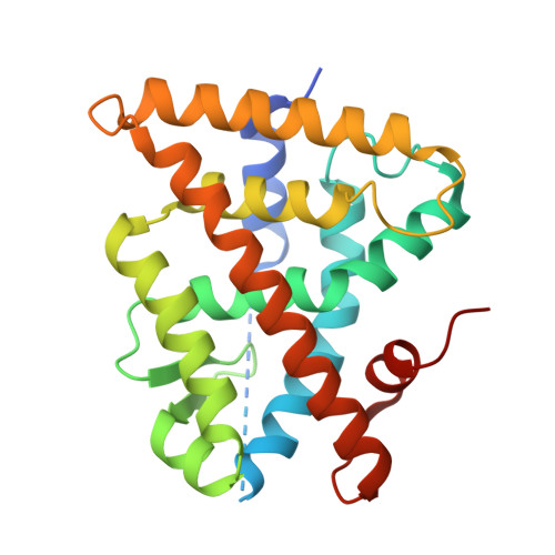 | |
UniProt & NIH Common Fund Data Resources | |||||
PHAROS: P19793 GTEx: ENSG00000186350 | |||||
Entity Groups | |||||
| Sequence Clusters | 30% Identity50% Identity70% Identity90% Identity95% Identity100% Identity | ||||
| UniProt Group | P19793 | ||||
Sequence AnnotationsExpand | |||||
Reference Sequence | |||||
Entity ID: 2 | |||||
|---|---|---|---|---|---|
| Molecule | Chains | Sequence Length | Organism | Details | Image |
| HIS-LYS-ILE-LEU-HIS-ARG-LEU-LEU-GLN | 12 | Homo sapiens | Mutation(s): 0 EC: 2.3.1.48 | 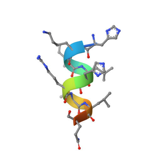 | |
UniProt & NIH Common Fund Data Resources | |||||
PHAROS: Q15788 GTEx: ENSG00000084676 | |||||
Entity Groups | |||||
| Sequence Clusters | 30% Identity50% Identity70% Identity90% Identity95% Identity100% Identity | ||||
| UniProt Group | Q15788 | ||||
Sequence AnnotationsExpand | |||||
Reference Sequence | |||||
| Ligands 1 Unique | |||||
|---|---|---|---|---|---|
| ID | Chains | Name / Formula / InChI Key | 2D Diagram | 3D Interactions | |
| WY5 (Subject of Investigation/LOI) Download:Ideal Coordinates CCD File | E [auth A], F [auth B] | 7-oxidanyl-2-oxidanylidene-6-(3,5,5,8,8-pentamethyl-6,7-dihydronaphthalen-2-yl)chromene-3-carboxylic acid C25 H26 O5 UYPOSIGCFZMVDD-UHFFFAOYSA-N |  | ||
| Length ( Å ) | Angle ( ˚ ) |
|---|---|
| a = 64.39 | α = 90 |
| b = 64.39 | β = 90 |
| c = 111.168 | γ = 90 |
| Software Name | Purpose |
|---|---|
| REFMAC | refinement |
| HKL-2000 | data reduction |
| SCALEPACK | data scaling |
| MOLREP | phasing |
| Funding Organization | Location | Grant Number |
|---|---|---|
| Japan Society for the Promotion of Science | Japan | 16K18688 |
| Japan Society for the Promotion of Science | Japan | 18K14391 |
| Japan Society for the Promotion of Science | Japan | 12J06716 |


