6JQR

Crystal structure of FLT3 in complex with gilteritinib


Experimental Data Snapshot

  • Method: X-RAY DIFFRACTION
  • Resolution: 2.20 Å
  • R-Value Free: 0.213 
  • R-Value Work: 0.179 
  • R-Value Observed: 0.181 

wwPDB Validation   3D Report Full Report


Ligand Structure Quality Assessment 


This is version 1.1 of the entry. See complete history


Literature

Effect of Fms-like tyrosine kinase 3 (FLT3) ligand (FL) on antitumor activity of gilteritinib, a FLT3 inhibitor, in mice xenografted with FL-overexpressing cells.

Kawase, T.Nakazawa, T.Eguchi, T.Tsuzuki, H.Ueno, Y.Amano, Y.Suzuki, T.Mori, M.Yoshida, T.

(2019) Oncotarget 10: 6111-6123

  • DOI: https://doi.org/10.18632/oncotarget.27222
  • Primary Citation of Related Structures:  
    6JQR

  • PubMed Abstract: 

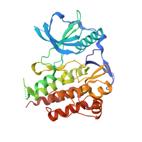
    Therapeutic effects of FLT3 inhibitors have been reported in acute myeloid leukemia (AML) with constitutively activating FLT3 mutations, including internal tandem duplication (ITD) and point mutation, which are found in approximately one-third of AML patients. One of the critical issues of treatment with FLT3 inhibitors in FLT3 -mutated AML is drug resistance. FLT3 ligand (FL) represents a mechanism of resistance to FLT3 inhibitors, including quizartinib, midostaurin, and sorafenib, in AML cells harboring both wild-type and mutant FLT3 ( FLT3 wt / FLT3 mut ). Here, we investigated the effect of FL on the efficacy of gilteritinib, a FLT3 inhibitor, in AML-derived cells in vitro and in mice. In contrast to other FLT3 inhibitors, FL stimulation had little effect on growth inhibition or apoptosis induction by gilteritinib. The antitumor activity of gilteritinib was also comparable between xenograft mouse models injected with FL-expressing and mock MOLM-13 cells. In the FLT3 signaling analyses, gilteritinib inhibited FLT3 wt and FLT3-ITD to a similar degree in HEK293 and Ba/F3 cells, and similarly suppressed FLT3 downstream signaling molecules (including ERK1/2 and STAT5) in both the presence and absence of FL in MOLM-13 cells. Co-crystal structure analysis showed that gilteritinib bound to the ATP-binding pocket of FLT3. These results suggest that gilteritinib has therapeutic potential in FLT3-mutated AML patients with FL overexpression.


  • Organizational Affiliation

    Drug Discovery Research, Astellas Pharma, Tsukuba-shi, Ibaraki, Japan.


Macromolecules
Find similar proteins by:  (by identity cutoff)  |  3D Structure
Entity ID: 1
MoleculeChains Sequence LengthOrganismDetailsImage
Receptor-type tyrosine-protein kinase FLT3,Receptor-type tyrosine-protein kinase FLT3330Homo sapiensMutation(s): 0 
EC: 2.7.10.1
UniProt & NIH Common Fund Data Resources
Find proteins for P36888 (Homo sapiens)
Explore P36888 
Go to UniProtKB:  P36888
PHAROS:  P36888
GTEx:  ENSG00000122025 
Entity Groups  
Sequence Clusters30% Identity50% Identity70% Identity90% Identity95% Identity100% Identity
UniProt GroupP36888
Sequence Annotations
Expand
  • Reference Sequence
Small Molecules
Ligands 4 Unique
IDChains Name / Formula / InChI Key2D Diagram3D Interactions
C6F
Query on C6F

Download Ideal Coordinates CCD File 
B [auth A]6-ethyl-3-[[3-methoxy-4-[4-(4-methylpiperazin-1-yl)piperidin-1-yl]phenyl]amino]-5-(oxan-4-ylamino)pyrazine-2-carboxamide
C29 H44 N8 O3
GYQYAJJFPNQOOW-UHFFFAOYSA-N
CXS
Query on CXS

Download Ideal Coordinates CCD File 
H [auth A]3-CYCLOHEXYL-1-PROPYLSULFONIC ACID
C9 H19 N O3 S
PJWWRFATQTVXHA-UHFFFAOYSA-N
SO4
Query on SO4

Download Ideal Coordinates CCD File 
C [auth A],
D [auth A],
E [auth A]
SULFATE ION
O4 S
QAOWNCQODCNURD-UHFFFAOYSA-L
GOL
Query on GOL

Download Ideal Coordinates CCD File 
F [auth A],
G [auth A],
I [auth A]
GLYCEROL
C3 H8 O3
PEDCQBHIVMGVHV-UHFFFAOYSA-N
Binding Affinity Annotations 
IDSourceBinding Affinity
C6F BindingDB:  6JQR IC50: min: 0.29, max: 0.41 (nM) from 2 assay(s)
Experimental Data & Validation

Experimental Data

  • Method: X-RAY DIFFRACTION
  • Resolution: 2.20 Å
  • R-Value Free: 0.213 
  • R-Value Work: 0.179 
  • R-Value Observed: 0.181 
  • Space Group: P 43 21 2
Unit Cell:
Length ( Å )Angle ( ˚ )
a = 82.367α = 90
b = 82.367β = 90
c = 146.469γ = 90
Software Package:
Software NamePurpose
REFMACrefinement
XDSdata reduction
XSCALEdata scaling

Structure Validation

View Full Validation Report


Ligand Structure Quality Assessment 


Entry History 

Deposition Data

  • Released Date: 2019-11-20 
  • Deposition Author(s): Amano, Y.

Revision History  (Full details and data files)

  • Version 1.0: 2019-11-20
    Type: Initial release
  • Version 1.1: 2024-03-27
    Changes: Data collection, Database references