6JXI

Rb+-bound E2-MgF state of the gastric proton pump (Tyr799Trp)


Experimental Data Snapshot

  • Method: X-RAY DIFFRACTION
  • Resolution: 2.60 Å
  • R-Value Free: 
    0.263 (Depositor), 0.260 (DCC) 
  • R-Value Work: 
    0.206 (Depositor), 0.210 (DCC) 
  • R-Value Observed: 
    0.209 (Depositor) 

Starting Model: experimental
View more details

wwPDB Validation   3D Report Full Report


Ligand Structure Quality Assessment 

Created with Raphaël 2.3.0Worse 01 BetterLigand structure goodness of fit to experimental dataBest fitted PCWClick on this verticalbar to view detailsBest fitted CE1Click on this verticalbar to view detailsBest fitted CLRClick on this verticalbar to view detailsBest fitted NAGClick on this verticalbar to view details

This is version 1.5 of the entry. See complete history


Literature

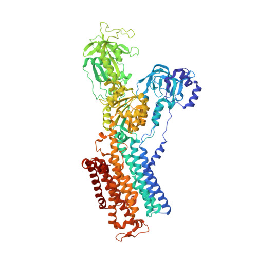
A single K + -binding site in the crystal structure of the gastric proton pump.

Yamamoto, K.Dubey, V.Irie, K.Nakanishi, H.Khandelia, H.Fujiyoshi, Y.Abe, K.

(2019) Elife 8

  • DOI: https://doi.org/10.7554/eLife.47701
  • Primary Citation of Related Structures:  
    6JXH, 6JXI, 6JXJ, 6JXK

  • PubMed Abstract: 

    The gastric proton pump (H + ,K + -ATPase), a P-type ATPase responsible for gastric acidification, mediates electro-neutral exchange of H + and K + coupled with ATP hydrolysis, but with an as yet undetermined transport stoichiometry. Here we show crystal structures at a resolution of 2.5 Å of the pump in the E2-P transition state, in which the counter-transporting cation is occluded. We found a single K + bound to the cation-binding site of the H + ,K + -ATPase, indicating an exchange of 1H + /1K + per hydrolysis of one ATP molecule. This fulfills the energy requirement for the generation of a six pH unit gradient across the membrane. The structural basis of K + recognition is resolved and supported by molecular dynamics simulations, establishing how the H + ,K + -ATPase overcomes the energetic challenge to generate an H + gradient of more than a million-fold-one of the highest cation gradients known in mammalian tissue-across the membrane.


  • Organizational Affiliation

    Cellular and Structural Physiology Institute, Nagoya University, Nagoya, Japan.


Macromolecules
Find similar proteins by:  (by identity cutoff)  |  3D Structure
Entity ID: 1
MoleculeChains Sequence LengthOrganismDetailsImage
Potassium-transporting ATPase alpha chain 1987Sus scrofaMutation(s): 4 
Gene Names: ATP4A
EC: 7.2.2.19
Membrane Entity: Yes 
UniProt
Find proteins for P19156 (Sus scrofa)
Explore P19156 
Go to UniProtKB:  P19156
Entity Groups  
Sequence Clusters30% Identity50% Identity70% Identity90% Identity95% Identity100% Identity
UniProt GroupP19156
Sequence Annotations
Expand
  • Reference Sequence
Find similar proteins by:  (by identity cutoff)  |  3D Structure
Entity ID: 2
MoleculeChains Sequence LengthOrganismDetailsImage
Potassium-transporting ATPase subunit beta289Sus scrofaMutation(s): 0 
Gene Names: ATP4B
Membrane Entity: Yes 
UniProt
Find proteins for P18434 (Sus scrofa)
Explore P18434 
Go to UniProtKB:  P18434
Entity Groups  
Sequence Clusters30% Identity50% Identity70% Identity90% Identity95% Identity100% Identity
UniProt GroupP18434
Glycosylation
Glycosylation Sites: 5
Sequence Annotations
Expand
  • Reference Sequence
Small Molecules
Ligands 7 Unique
IDChains Name / Formula / InChI Key2D Diagram3D Interactions
PCW
Query on PCW

Download Ideal Coordinates CCD File 
D [auth A],
F [auth A],
N [auth A],
Q [auth B]
1,2-DIOLEOYL-SN-GLYCERO-3-PHOSPHOCHOLINE
C44 H85 N O8 P
SNKAWJBJQDLSFF-NVKMUCNASA-O
CE1
Query on CE1

Download Ideal Coordinates CCD File 
E [auth A]O-DODECANYL OCTAETHYLENE GLYCOL
C28 H58 O9
YYELLDKEOUKVIQ-UHFFFAOYSA-N
CLR
Query on CLR

Download Ideal Coordinates CCD File 
R [auth B]CHOLESTEROL
C27 H46 O
HVYWMOMLDIMFJA-DPAQBDIFSA-N
NAG
Query on NAG

Download Ideal Coordinates CCD File 
O [auth B],
P [auth B],
S [auth B],
T [auth B],
U [auth B]
2-acetamido-2-deoxy-beta-D-glucopyranose
C8 H15 N O6
OVRNDRQMDRJTHS-FMDGEEDCSA-N
MF4
Query on MF4

Download Ideal Coordinates CCD File 
C [auth A]TETRAFLUOROMAGNESATE(2-)
F4 Mg
XVYWAXYEHHUKQW-UHFFFAOYSA-J
RB
Query on RB

Download Ideal Coordinates CCD File 
H [auth A]
I [auth A]
J [auth A]
K [auth A]
L [auth A]
RUBIDIUM ION
Rb
NCCSSGKUIKYAJD-UHFFFAOYSA-N
MG
Query on MG

Download Ideal Coordinates CCD File 
G [auth A]MAGNESIUM ION
Mg
JLVVSXFLKOJNIY-UHFFFAOYSA-N
Experimental Data & Validation

Experimental Data

  • Method: X-RAY DIFFRACTION
  • Resolution: 2.60 Å
  • R-Value Free:  0.263 (Depositor), 0.260 (DCC) 
  • R-Value Work:  0.206 (Depositor), 0.210 (DCC) 
  • R-Value Observed: 0.209 (Depositor) 
Space Group: P 31 2 1
Unit Cell:
Length ( Å )Angle ( ˚ )
a = 103.38α = 90
b = 103.38β = 90
c = 369.86γ = 120
Software Package:
Software NamePurpose
PHENIXrefinement
PHENIXrefinement
XDSdata reduction
XDSdata scaling
PHASERphasing

Structure Validation

View Full Validation Report


Ligand Structure Quality Assessment 

Created with Raphaël 2.3.0Worse 01 BetterLigand structure goodness of fit to experimental dataBest fitted PCWClick on this verticalbar to view detailsBest fitted CE1Click on this verticalbar to view detailsBest fitted CLRClick on this verticalbar to view detailsBest fitted NAGClick on this verticalbar to view details

Entry History 

Deposition Data

Revision History  (Full details and data files)

  • Version 1.0: 2019-08-14
    Type: Initial release
  • Version 1.1: 2019-09-04
    Changes: Data collection, Database references
  • Version 1.2: 2020-07-29
    Type: Remediation
    Reason: Carbohydrate remediation
    Changes: Data collection, Derived calculations, Structure summary
  • Version 1.3: 2020-09-30
    Changes: Database references, Structure summary
  • Version 1.4: 2023-11-22
    Changes: Data collection, Database references, Refinement description
  • Version 1.5: 2024-11-13
    Changes: Structure summary