


Crystal structure of dimeric RXRalpha-LBD complexed with partial agonist CBt-PMN and SRC1
Shimizu, K., Numoto, N., Nakano, S., Makishima, M., Kakuta, H., Ito, N.To be published.
Experimental Data Snapshot
Starting Model: experimental
View more details
Entity ID: 1 | |||||
|---|---|---|---|---|---|
| Molecule | Chains | Sequence Length | Organism | Details | Image |
| Retinoic acid receptor RXR-alpha | 243 | Homo sapiens | Mutation(s): 0 Gene Names: RXRA, NR2B1 | 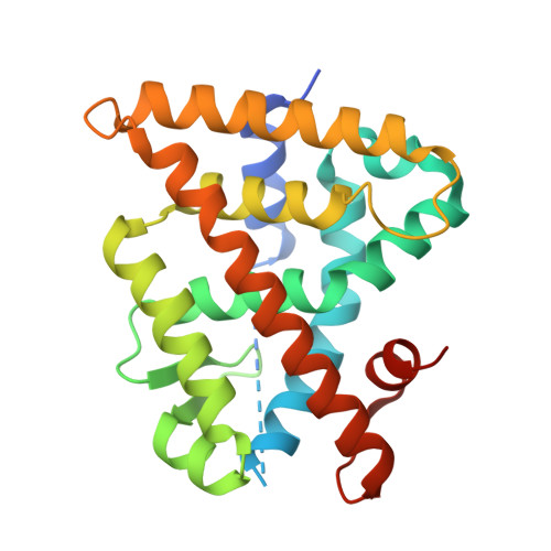 | |
UniProt & NIH Common Fund Data Resources | |||||
PHAROS: P19793 GTEx: ENSG00000186350 | |||||
Entity Groups | |||||
| Sequence Clusters | 30% Identity50% Identity70% Identity90% Identity95% Identity100% Identity | ||||
| UniProt Group | P19793 | ||||
Sequence AnnotationsExpand | |||||
Reference Sequence | |||||
Entity ID: 2 | |||||
|---|---|---|---|---|---|
| Molecule | Chains | Sequence Length | Organism | Details | Image |
| Nuclear receptor coactivator 1 | 12 | Homo sapiens | Mutation(s): 0 EC: 2.3.1.48 | 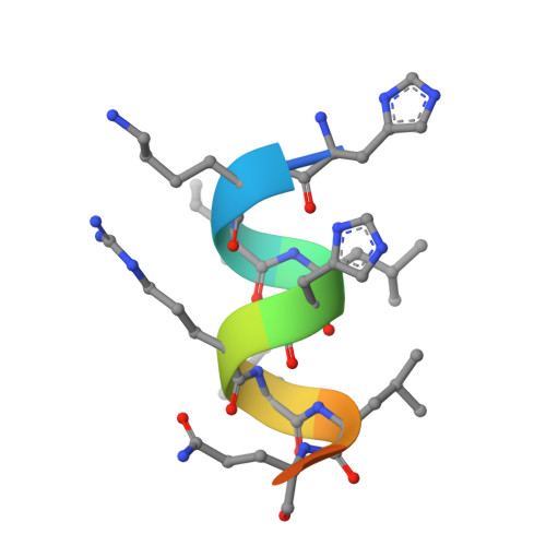 | |
UniProt & NIH Common Fund Data Resources | |||||
PHAROS: Q15788 GTEx: ENSG00000084676 | |||||
Entity Groups | |||||
| Sequence Clusters | 30% Identity50% Identity70% Identity90% Identity95% Identity100% Identity | ||||
| UniProt Group | Q15788 | ||||
Sequence AnnotationsExpand | |||||
Reference Sequence | |||||
| Ligands 2 Unique | |||||
|---|---|---|---|---|---|
| ID | Chains | Name / Formula / InChI Key | 2D Diagram | 3D Interactions | |
| 9HF (Subject of Investigation/LOI) Download:Ideal Coordinates CCD File | C [auth A] | 1-(3,5,5,8,8-pentamethyl-6,7-dihydronaphthalen-2-yl)benzotriazole-5-carboxylic acid C22 H25 N3 O2 QSRQAKVNYDAVIT-UHFFFAOYSA-N |  | ||
| CA Download:Ideal Coordinates CCD File | D [auth A] | CALCIUM ION Ca BHPQYMZQTOCNFJ-UHFFFAOYSA-N |  | ||
| Length ( Å ) | Angle ( ˚ ) |
|---|---|
| a = 67.837 | α = 90 |
| b = 67.837 | β = 90 |
| c = 105.153 | γ = 90 |
| Software Name | Purpose |
|---|---|
| PHENIX | refinement |
| XDS | data reduction |
| XDS | data scaling |
| PHASER | phasing |


