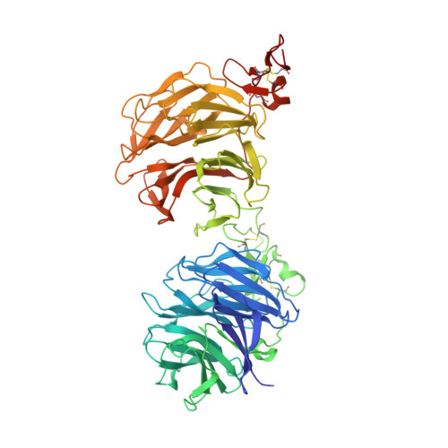
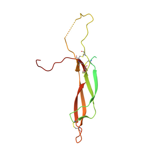
6L6R

Crystal structure of LRP6 E1E2-SOST complex


Experimental Data Snapshot

  • Method: X-RAY DIFFRACTION
  • Resolution: 3.80 Å
  • R-Value Free: 0.256 
  • R-Value Work: 0.207 
  • R-Value Observed: 0.211 

Starting Models: experimental
View more details

wwPDB Validation   3D Report Full Report


Ligand Structure Quality Assessment 


This is version 1.3 of the entry. See complete history


Literature

Sclerostin inhibits Wnt signaling through tandem interaction with two LRP6 ectodomains.

Kim, J.Han, W.Park, T.Kim, E.J.Bang, I.Lee, H.S.Jeong, Y.Roh, K.Kim, J.Kim, J.S.Kang, C.Seok, C.Han, J.-K.Choi, H.-J.

(2020) Nat Commun 11: 5357-5357

  • DOI: https://doi.org/10.1038/s41467-020-19155-4
  • Primary Citation of Related Structures:  
    6L6R

  • PubMed Abstract: 

    Low-density lipoprotein receptor-related protein 6 (LRP6) is a coreceptor of the β-catenin-dependent Wnt signaling pathway. The LRP6 ectodomain binds Wnt proteins, as well as Wnt inhibitors such as sclerostin (SOST), which negatively regulates Wnt signaling in osteocytes. Although LRP6 ectodomain 1 (E1) is known to interact with SOST, several unresolved questions remain, such as the reason why SOST binds to LRP6 E1E2 with higher affinity than to the E1 domain alone. Here, we present the crystal structure of the LRP6 E1E2-SOST complex with two interaction sites in tandem. The unexpected additional binding site was identified between the C-terminus of SOST and the LRP6 E2 domain. This interaction was confirmed by in vitro binding and cell-based signaling assays. Its functional significance was further demonstrated in vivo using Xenopus laevis embryos. Our results provide insights into the inhibitory mechanism of SOST on Wnt signaling.


  • Organizational Affiliation

    Department of Biological Sciences, Seoul National University, Seoul, 08826, Republic of Korea.


Macromolecules
Find similar proteins by:  (by identity cutoff)  |  3D Structure
Entity ID: 1
MoleculeChains Sequence LengthOrganismDetailsImage
Low-density lipoprotein receptor-related protein 6
A, B
622Homo sapiensMutation(s): 0 
Gene Names: LRP6
UniProt & NIH Common Fund Data Resources
Find proteins for O75581 (Homo sapiens)
Explore O75581 
Go to UniProtKB:  O75581
PHAROS:  O75581
GTEx:  ENSG00000070018 
Entity Groups  
Sequence Clusters30% Identity50% Identity70% Identity90% Identity95% Identity100% Identity
UniProt GroupO75581
Glycosylation
Glycosylation Sites: 3Go to GlyGen: O75581-1
Sequence Annotations
Expand
  • Reference Sequence
Find similar proteins by:  (by identity cutoff)  |  3D Structure
Entity ID: 2
MoleculeChains Sequence LengthOrganismDetailsImage
Sclerostin
C, D
158Homo sapiensMutation(s): 0 
Gene Names: SOSTUNQ2976/PRO7455/PRO7476
UniProt & NIH Common Fund Data Resources
Find proteins for Q9BQB4 (Homo sapiens)
Explore Q9BQB4 
Go to UniProtKB:  Q9BQB4
PHAROS:  Q9BQB4
GTEx:  ENSG00000167941 
Entity Groups  
Sequence Clusters30% Identity50% Identity70% Identity90% Identity95% Identity100% Identity
UniProt GroupQ9BQB4
Sequence Annotations
Expand
  • Reference Sequence
Oligosaccharides

Help

Entity ID: 3
MoleculeChains Length2D Diagram Glycosylation3D Interactions
2-acetamido-2-deoxy-beta-D-glucopyranose-(1-4)-2-acetamido-2-deoxy-beta-D-glucopyranose
E, G
2N-Glycosylation
Glycosylation Resources
GlyTouCan:  G42666HT
GlyCosmos:  G42666HT
GlyGen:  G42666HT
Entity ID: 4
MoleculeChains Length2D Diagram Glycosylation3D Interactions
alpha-L-fucopyranose-(1-4)-[alpha-L-fucopyranose-(1-6)]2-acetamido-2-deoxy-beta-D-glucopyranose
F, H
3N-Glycosylation
Glycosylation Resources
GlyTouCan:  G99664XK
GlyCosmos:  G99664XK
GlyGen:  G99664XK
Small Molecules
Ligands 3 Unique
IDChains Name / Formula / InChI Key2D Diagram3D Interactions
NAG
Query on NAG

Download Ideal Coordinates CCD File 
I [auth A],
M [auth B]
2-acetamido-2-deoxy-beta-D-glucopyranose
C8 H15 N O6
OVRNDRQMDRJTHS-FMDGEEDCSA-N
ACT
Query on ACT

Download Ideal Coordinates CCD File 
L [auth A]ACETATE ION
C2 H3 O2
QTBSBXVTEAMEQO-UHFFFAOYSA-M
MG
Query on MG

Download Ideal Coordinates CCD File 
J [auth A],
K [auth A],
N [auth B],
O [auth B]
MAGNESIUM ION
Mg
JLVVSXFLKOJNIY-UHFFFAOYSA-N
Experimental Data & Validation

Experimental Data

  • Method: X-RAY DIFFRACTION
  • Resolution: 3.80 Å
  • R-Value Free: 0.256 
  • R-Value Work: 0.207 
  • R-Value Observed: 0.211 
  • Space Group: P 64
Unit Cell:
Length ( Å )Angle ( ˚ )
a = 252.792α = 90
b = 252.792β = 90
c = 86.286γ = 120
Software Package:
Software NamePurpose
PHENIXrefinement
XDSdata reduction
Aimlessdata scaling
PHASERphasing
PDB_EXTRACTdata extraction

Structure Validation

View Full Validation Report


Ligand Structure Quality Assessment 


Entry History & Funding Information

Deposition Data


Funding OrganizationLocationGrant Number
National Research Foundation (NRF, Korea)Korea, Republic OfNRF-2016R1A2B4013488
National Research Foundation (NRF, Korea)Korea, Republic OfNRF-2019M3E5D6063903

Revision History  (Full details and data files)

  • Version 1.0: 2020-10-28
    Type: Initial release
  • Version 1.1: 2020-11-04
    Changes: Database references
  • Version 1.2: 2023-11-22
    Changes: Data collection, Database references, Refinement description
  • Version 1.3: 2024-11-13
    Changes: Structure summary