6L93 | pdb_00006l93

X-ray structure of the ligand-free human TRPV1 ankyrin repeat domain


Experimental Data Snapshot

  • Method: X-RAY DIFFRACTION
  • Resolution: 4.47 Å
  • R-Value Free: 
    0.270 (Depositor), 0.272 (DCC) 
  • R-Value Work: 
    0.220 (Depositor), 0.225 (DCC) 
  • R-Value Observed: 
    0.223 (Depositor) 

Starting Model: experimental
View more details

wwPDB Validation 3D Report Full Report

Validation slider image for 6L93

This is version 1.1 of the entry. See complete history

Literature

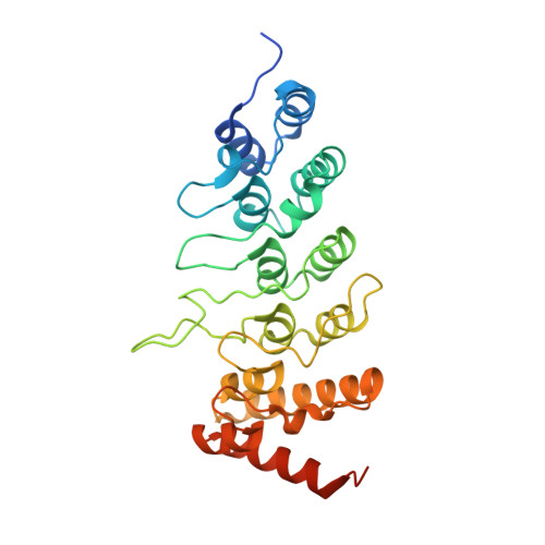
Structure determination of the human TRPV1 ankyrin-repeat domain under nonreducing conditions.

Tanaka, M.Hayakawa, K.Ogawa, N.Kurokawa, T.Kitanishi, K.Ite, K.Matsui, T.Mori, Y.Unno, M.

(2020) Acta Crystallogr F Struct Biol Commun 76: 130-137

  • DOI: https://doi.org/10.1107/S2053230X20001533
  • Primary Citation Related Structures: 
    6L93

  • PubMed Abstract: 

    TRPV1, a member of the transient receptor potential (TRP) channels family, has been found to be involved in redox sensing. The crystal structure of the human TRPV1 ankyrin-repeat domain (TRPV1-ARD) was determined at 4.5 Å resolution under nonreducing conditions. This is the first report of the crystal structure of a ligand-free form of TRPV1-ARD and in particular of the human homologue. The structure showed a unique conformation in finger loop 3 near Cys258, which is most likely to be involved in inter-subunit disulfide-bond formation. Also, in human TRPV1-ARD it was possible for solvent to access Cys258. This structural feature might be related to the high sensitivity of human TRPV1 to oxidants. ESI-MS revealed that Cys258 did not form an S-OH functionality even under nonreducing conditions.


  • Organizational Affiliation
    • Graduate School of Science and Engineering, Ibaraki University, Hitachi, Ibaraki 316-8511, Japan.

Macromolecule Content 

  • Total Structure Weight: 185 kDa 
  • Atom Count: 11,813 
  • Modeled Residue Count: 1,518 
  • Deposited Residue Count: 1,680 
  • Unique protein chains: 1

Macromolecules

Find similar proteins by:|  3D Structure
Entity ID: 1
MoleculeChains  Sequence LengthOrganismDetailsImage
Transient receptor potential cation channel subfamily V member 1
A, B, C, D, E
A, B, C, D, E, F
280Homo sapiensMutation(s): 0 
Gene Names: TRPV1VR1
UniProt & NIH Common Fund Data Resources
Find proteins for Q8NER1 (Homo sapiens)
Explore Q8NER1 
Go to UniProtKB:  Q8NER1
PHAROS:  Q8NER1
GTEx:  ENSG00000196689 
Entity Groups
Sequence Clusters30% Identity50% Identity70% Identity90% Identity95% Identity100% Identity
UniProt GroupQ8NER1
Sequence Annotations
Expand
Reference Sequence

Experimental Data & Validation

Experimental Data

  • Method: X-RAY DIFFRACTION
  • Resolution: 4.47 Å
  • R-Value Free:  0.270 (Depositor), 0.272 (DCC) 
  • R-Value Work:  0.220 (Depositor), 0.225 (DCC) 
  • R-Value Observed: 0.223 (Depositor) 
Space Group: C 1 2 1
Unit Cell:
Length ( Å )Angle ( ˚ )
a = 184.228α = 90
b = 167.414β = 108.375
c = 112.632γ = 90
Software Package:
Software NamePurpose
PHENIXrefinement
PHENIXrefinement
XDSdata reduction
Aimlessdata scaling
PHASERphasing

Structure Validation

View Full Validation Report


Entry History 

Deposition Data

Revision History  (Full details and data files)

  • Version 1.0: 2020-03-18
    Type: Initial release
  • Version 1.1: 2023-11-22
    Changes: Data collection, Database references, Refinement description