6L96

Structure of PPARalpha-LBD/pemafibrate/SRC1 peptide


Experimental Data Snapshot

  • Method: X-RAY DIFFRACTION
  • Resolution: 3.20 Å
  • R-Value Free: 0.253 
  • R-Value Work: 0.187 
  • R-Value Observed: 0.190 

wwPDB Validation   3D Report Full Report


Ligand Structure Quality Assessment 


This is version 1.2 of the entry. See complete history


Literature

Elucidation of Molecular Mechanism of a Selective PPAR alpha Modulator, Pemafibrate, through Combinational Approaches of X-ray Crystallography, Thermodynamic Analysis, and First-Principle Calculations.

Kawasaki, M.Kambe, A.Yamamoto, Y.Arulmozhiraja, S.Ito, S.Nakagawa, Y.Tokiwa, H.Nakano, S.Shimano, H.

(2020) Int J Mol Sci 21

  • DOI: https://doi.org/10.3390/ijms21010361
  • Primary Citation of Related Structures:  
    6L96

  • PubMed Abstract: 

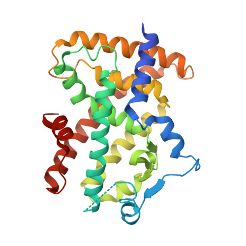
    The selective PPARα modulator (SPPARMα) is expected to medicate dyslipidemia with minimizing adverse effects. Recently, pemafibrate was screened from the ligand library as an SPPARMα bearing strong potency. Several clinical pieces of evidence have proved the usefulness of pemafibrate as a medication; however, how pemafibrate works as a SPPARMα at the molecular level is not fully known. In this study, we investigate the molecular mechanism behind its novel SPPARMα character through a combination of approaches of X-ray crystallography, isothermal titration calorimetry (ITC), and fragment molecular orbital (FMO) analysis. ITC measurements have indicated that pemafibrate binds more strongly to PPARα than to PPARγ. The crystal structure of PPARα-ligand binding domain (LBD)/pemafibrate/steroid receptor coactivator-1 peptide (SRC1) determined at 3.2 Å resolution indicates that pemafibrate binds to the ligand binding pocket (LBP) of PPARα in a Y-shaped form. The structure also reveals that the conformation of the phenoxyalkyl group in pemafibrate is flexible in the absence of SRC1 coactivator peptide bound to PPARα; this gives a freedom for the phenoxyalkyl group to adopt structural changes induced by the binding of coactivators. FMO calculations have indicated that the accumulation of hydrophobic interactions provided by the residues at the LBP improve the interaction between pemafibrate and PPARα compared with the interaction between fenofibrate and PPARα.


  • Organizational Affiliation

    Graduate Division of Nutritional and Environmental Sciences, University of Shizuoka, 52-1 Yada, Suruga-ku, Shizuoka 422-8526, Japan.


Macromolecules
Find similar proteins by:  (by identity cutoff)  |  3D Structure
Entity ID: 1
MoleculeChains Sequence LengthOrganismDetailsImage
Peroxisome proliferator-activated receptor alpha
A, B
275Homo sapiensMutation(s): 0 
Gene Names: PPARANR1C1PPAR
UniProt & NIH Common Fund Data Resources
Find proteins for Q07869 (Homo sapiens)
Explore Q07869 
Go to UniProtKB:  Q07869
PHAROS:  Q07869
GTEx:  ENSG00000186951 
Entity Groups  
Sequence Clusters30% Identity50% Identity70% Identity90% Identity95% Identity100% Identity
UniProt GroupQ07869
Sequence Annotations
Expand
  • Reference Sequence

Find similar proteins by:  Sequence   |   3D Structure  

Entity ID: 2
MoleculeChains Sequence LengthOrganismDetailsImage
SRC1 coactivator peptide13Homo sapiensMutation(s): 0 
UniProt & NIH Common Fund Data Resources
Find proteins for Q15788 (Homo sapiens)
Explore Q15788 
Go to UniProtKB:  Q15788
PHAROS:  Q15788
GTEx:  ENSG00000084676 
Entity Groups  
Sequence Clusters30% Identity50% Identity70% Identity90% Identity95% Identity100% Identity
UniProt GroupQ15788
Sequence Annotations
Expand
  • Reference Sequence
Small Molecules
Ligands 1 Unique
IDChains Name / Formula / InChI Key2D Diagram3D Interactions
P7F (Subject of Investigation/LOI)
Query on P7F

Download Ideal Coordinates CCD File 
D [auth A],
E [auth B]
(2~{R})-2-[3-[[1,3-benzoxazol-2-yl-[3-(4-methoxyphenoxy)propyl]amino]methyl]phenoxy]butanoic acid
C28 H30 N2 O6
ZHKNLJLMDFQVHJ-RUZDIDTESA-N
Binding Affinity Annotations 
IDSourceBinding Affinity
P7F BindingDB:  6L96 EC50: 1 (nM) from 1 assay(s)
Experimental Data & Validation

Experimental Data

  • Method: X-RAY DIFFRACTION
  • Resolution: 3.20 Å
  • R-Value Free: 0.253 
  • R-Value Work: 0.187 
  • R-Value Observed: 0.190 
  • Space Group: P 31 2 1
Unit Cell:
Length ( Å )Angle ( ˚ )
a = 82.736α = 90
b = 82.736β = 90
c = 177.521γ = 120
Software Package:
Software NamePurpose
REFMACrefinement
HKL-2000data reduction
SCALEPACKdata scaling
MOLREPphasing

Structure Validation

View Full Validation Report


Ligand Structure Quality Assessment 


Entry History & Funding Information

Deposition Data


Funding OrganizationLocationGrant Number
Japan Society for the Promotion of ScienceJapan18K14391
Japan Society for the Promotion of ScienceJapan16K18688

Revision History  (Full details and data files)

  • Version 1.0: 2020-01-15
    Type: Initial release
  • Version 1.1: 2020-01-29
    Changes: Database references
  • Version 1.2: 2023-11-22
    Changes: Data collection, Database references, Refinement description