6M4W

Crystal structure of MBP fused split FKBP-FRB T2098L mutant in complex with rapamycin


Experimental Data Snapshot

  • Method: X-RAY DIFFRACTION
  • Resolution: 3.11 Å
  • R-Value Free: 0.278 
  • R-Value Work: 0.236 
  • R-Value Observed: 0.238 

wwPDB Validation   3D Report Full Report


Ligand Structure Quality Assessment 


This is version 1.2 of the entry. See complete history


Literature

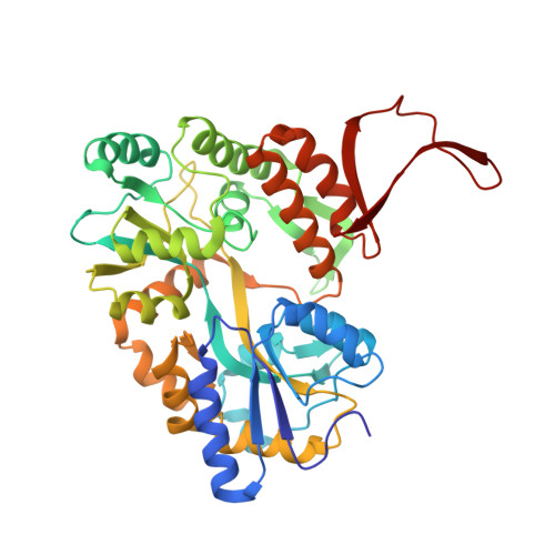
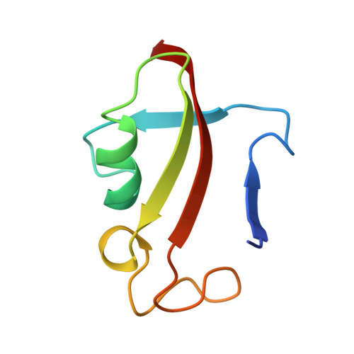
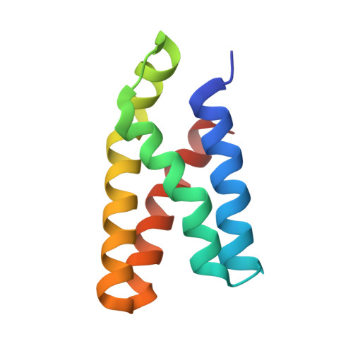
Rational design and implementation of a chemically inducible heterotrimerization system.

Wu, H.D.Kikuchi, M.Dagliyan, O.Aragaki, A.K.Nakamura, H.Dokholyan, N.V.Umehara, T.Inoue, T.

(2020) Nat Methods 17: 928-936

  • DOI: https://doi.org/10.1038/s41592-020-0913-x
  • Primary Citation of Related Structures:  
    6M4U, 6M4V, 6M4W

  • PubMed Abstract: 

    Chemically inducible dimerization (CID) uses a small molecule to induce binding of two different proteins. CID tools such as the FK506-binding protein-FKBP-rapamycin-binding- (FKBP-FRB)-rapamycin system have been widely used to probe molecular events inside and outside cells. While various CID tools are available, chemically inducible trimerization (CIT) does not exist, due to inherent challenges in designing a chemical that simultaneously binds three proteins with high affinity and specificity. Here, we developed CIT by rationally splitting FRB and FKBP. Cellular and structural datasets showed efficient trimerization of split pairs of FRB or FKBP with full-length FKBP or FRB, respectively, by rapamycin. CIT rapidly induced tri-organellar junctions and perturbed intended membrane lipids exclusively at select membrane contact sites. By conferring one additional condition to what is achievable with CID, CIT expands the types of manipulation in single live cells to address cell biology questions otherwise intractable and engineer cell functions for future synthetic biology applications.


  • Organizational Affiliation

    Department of Biomedical Engineering, Johns Hopkins University School of Medicine, Baltimore, MD, USA.


Macromolecules
Find similar proteins by:  (by identity cutoff)  |  3D Structure
Entity ID: 1
MoleculeChains Sequence LengthOrganismDetailsImage
chimera of Maltose/maltodextrin-binding periplasmic protein and Peptidyl-prolyl cis-trans isomerase FKBP1A
A, B, C
405Escherichia coli K-12Homo sapiens
This entity is chimeric
Mutation(s): 5 
Gene Names: malEb4034JW3994FKBP1AFKBP1FKBP12
EC: 5.2.1.8
UniProt & NIH Common Fund Data Resources
Find proteins for P0AEX9 (Escherichia coli (strain K12))
Explore P0AEX9 
Go to UniProtKB:  P0AEX9
Find proteins for P62942 (Homo sapiens)
Explore P62942 
Go to UniProtKB:  P62942
PHAROS:  P62942
GTEx:  ENSG00000088832 
Entity Groups  
Sequence Clusters30% Identity50% Identity70% Identity90% Identity95% Identity100% Identity
UniProt GroupsP62942P0AEX9
Sequence Annotations
Expand
  • Reference Sequence
Find similar proteins by:  (by identity cutoff)  |  3D Structure
Entity ID: 2
MoleculeChains Sequence LengthOrganismDetailsImage
Peptidyl-prolyl cis-trans isomerase FKBP1A
D, E, F
76Homo sapiensMutation(s): 0 
Gene Names: FKBP1AFKBP1FKBP12
EC: 5.2.1.8
UniProt & NIH Common Fund Data Resources
Find proteins for P62942 (Homo sapiens)
Explore P62942 
Go to UniProtKB:  P62942
PHAROS:  P62942
GTEx:  ENSG00000088832 
Entity Groups  
Sequence Clusters30% Identity50% Identity70% Identity90% Identity95% Identity100% Identity
UniProt GroupP62942
Sequence Annotations
Expand
  • Reference Sequence
Find similar proteins by:  (by identity cutoff)  |  3D Structure
Entity ID: 3
MoleculeChains Sequence LengthOrganismDetailsImage
Serine/threonine-protein kinase mTOR
G, H, I
95Homo sapiensMutation(s): 1 
Gene Names: MTOR
EC: 2.7.11.1
UniProt & NIH Common Fund Data Resources
Find proteins for P42345 (Homo sapiens)
Explore P42345 
Go to UniProtKB:  P42345
PHAROS:  P42345
GTEx:  ENSG00000198793 
Entity Groups  
Sequence Clusters30% Identity50% Identity70% Identity90% Identity95% Identity100% Identity
UniProt GroupP42345
Sequence Annotations
Expand
  • Reference Sequence
Oligosaccharides

Help

Entity ID: 4
MoleculeChains Length2D Diagram Glycosylation3D Interactions
alpha-D-glucopyranose-(1-4)-alpha-D-glucopyranose
J, K, L
2N/A
Glycosylation Resources
GlyTouCan:  G07411ON
GlyCosmos:  G07411ON
Biologically Interesting Molecules (External Reference) 1 Unique
Experimental Data & Validation

Experimental Data

  • Method: X-RAY DIFFRACTION
  • Resolution: 3.11 Å
  • R-Value Free: 0.278 
  • R-Value Work: 0.236 
  • R-Value Observed: 0.238 
  • Space Group: P 43 21 2
Unit Cell:
Length ( Å )Angle ( ˚ )
a = 127.546α = 90
b = 127.546β = 90
c = 278.557γ = 90
Software Package:
Software NamePurpose
XDSdata reduction
MOLREPphasing
REFMACrefinement
Aimlessdata scaling

Structure Validation

View Full Validation Report


Ligand Structure Quality Assessment 


Entry History 

Deposition Data

Revision History  (Full details and data files)

  • Version 1.0: 2020-08-26
    Type: Initial release
  • Version 1.1: 2020-09-16
    Changes: Database references
  • Version 1.2: 2023-11-29
    Changes: Data collection, Database references, Refinement description