


Crystal structure of Tdp1 catalytic domain
Lountos, G.T., Zhao, X.Z., Kiselev, E., Tropea, J.E., Needle, D., Burke Jr., T.R., Pommier, Y., Waugh, D.S.To be published.
Experimental Data Snapshot
Starting Model: experimental
View more details
Entity ID: 1 | |||||
|---|---|---|---|---|---|
| Molecule | Chains | Sequence Length | Organism | Details | Image |
| Tyrosyl-DNA phosphodiesterase 1 | 461 | Homo sapiens | Mutation(s): 0 Gene Names: TDP1 EC: 3.1.4 | 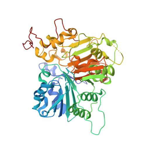 | |
UniProt & NIH Common Fund Data Resources | |||||
PHAROS: Q9NUW8 GTEx: ENSG00000042088 | |||||
Entity Groups | |||||
| Sequence Clusters | 30% Identity50% Identity70% Identity90% Identity95% Identity100% Identity | ||||
| UniProt Group | Q9NUW8 | ||||
Sequence AnnotationsExpand | |||||
Reference Sequence | |||||
| Ligands 3 Unique | |||||
|---|---|---|---|---|---|
| ID | Chains | Name / Formula / InChI Key | 2D Diagram | 3D Interactions | |
| K8D Download:Ideal Coordinates CCD File | C [auth A], H [auth B] | 4-(methylamino)benzene-1,2-dicarboxylic acid C9 H9 N O4 WIVUEBNYALSDOC-UHFFFAOYSA-N |  | ||
| DMS Download:Ideal Coordinates CCD File | G [auth A] | DIMETHYL SULFOXIDE C2 H6 O S IAZDPXIOMUYVGZ-UHFFFAOYSA-N |  | ||
| EDO Download:Ideal Coordinates CCD File | D [auth A], E [auth A], F [auth A], I [auth B] | 1,2-ETHANEDIOL C2 H6 O2 LYCAIKOWRPUZTN-UHFFFAOYSA-N |  | ||
| Length ( Å ) | Angle ( ˚ ) |
|---|---|
| a = 50.043 | α = 90 |
| b = 105.399 | β = 90 |
| c = 193.59 | γ = 90 |
| Software Name | Purpose |
|---|---|
| PHENIX | refinement |
| HKL-3000 | data reduction |
| HKL-3000 | data scaling |
| PHASER | phasing |


