A Novel Class of Common Docking Domain Inhibitors That Prevent ERK2 Activation and Substrate Phosphorylation.
Sammons, R.M., Perry, N.A., Li, Y., Cho, E.J., Piserchio, A., Zamora-Olivares, D.P., Ghose, R., Kaoud, T.S., Debevec, G., Bartholomeusz, C., Gurevich, V.V., Iverson, T.M., Giulianotti, M., Houghten, R.A., Dalby, K.N.(2019) ACS Chem Biol 14: 1183-1194
- PubMed: 31058487 Search on PubMedSearch on PubMed Central
- DOI: https://doi.org/10.1021/acschembio.9b00093
- Primary Citation Related Structures:
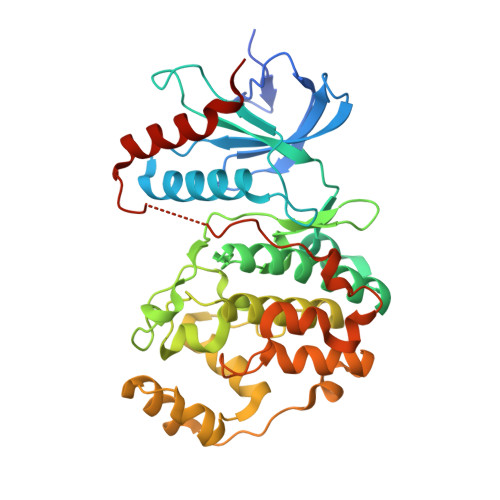
6NBS - PubMed Abstract:
Extracellular signal-regulated kinases (ERK1/2) are mitogen-activated protein kinases (MAPKs) that play a pro-tumorigenic role in numerous cancers. ERK1/2 possess two protein-docking sites that are distinct from the active site: the D-recruitment site (DRS) and the F-recruitment site. These docking sites facilitate substrate recognition, intracellular localization, signaling specificity, and protein complex assembly. Targeting these sites on ERK in a therapeutic context may overcome many problems associated with traditional ATP-competitive inhibitors. Here, we identified a new class of inhibitors that target the ERK DRS by screening a synthetic combinatorial library of more than 30 million compounds. The screen detects the competitive displacement of a fluorescent peptide from the DRS of ERK2. The top molecular scaffold from the screen was optimized for structure-activity relationship by positional scanning of different functional groups. This resulted in 10 compounds with similar binding affinities and a shared core structure consisting of a tertiary amine hub with three functionalized cyclic guanidino branches. Compound 2507-1 inhibited ERK2 from phosphorylating a DRS-targeting substrate and prevented the phosphorylation of ERK2 by a constitutively active MEK1 (MAPK/ERK kinase 1) mutant. Interaction between an analogue, 2507-8, and the ERK2 DRS was confirmed by nuclear magnetic resonance and X-ray crystallography. 2507-8 forms critical interactions at the common docking domain residue Asp319 via an arginine-like moiety that is shared by all 10 hits, suggesting a common binding mode. The structural and biochemical insights reported here provide the basis for developing new ERK inhibitors that are not ATP-competitive but instead function by disrupting critical protein-protein interactions.
- Torrey Pines Institute for Molecular Studies , Port St. Lucie , Florida 34987 , United States.
Organizational Affiliation: