Beyond Traditional Structure-Based Drug Design: The Role of Iron Complexation, Strain, and Water in the Binding of Inhibitors for Hypoxia-Inducible Factor Prolyl Hydroxylase 2.
Bembenek, S.D., Venkatesan, H., Peltier, H.M., Rosen, M.D., Barrett, T.D., Kanelakis, K.C., Palomino, H.L., Brondstetter, T.I., Mirzadegan, T., Rabinowitz, M.H.(2019) ACS Omega 4: 6703-6708
- PubMed: 31179408 Search on PubMedSearch on PubMed Central
- DOI: https://doi.org/10.1021/acsomega.9b00199
- Primary Citation Related Structures:
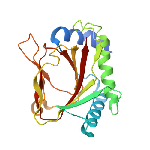
6NMQ - PubMed Abstract:
A combination of structure-based drug design and medicinal chemistry efforts led us from benzimidazole-2-carboxamide with modestly active hypoxia-inducible factor prolyl hydroxylase 2 inhibition to certain benzimidazole-2-pyrazole carboxylic acids that were more potent as well as orally efficacious stimulators of erythropoietin secretion in our in vivo mouse model. To better understand the structure-activity relationship, it was necessary to account for (i) the complexation of the ligand with the active site Fe 2+ , (ii) the strain incurred by the ligand upon binding, and (iii) certain key water interactions identified by a crystal structure analysis. With this more complete computational model, we arrived at an overarching paradigm that accounted for the potency differences between benzimidazole-2-carboxamide and benzimidazole-2-pyrazole carboxylic acid enzyme inhibitors. Moreover, the computational paradigm allowed us to anticipate that the bioisostere replacement strategy (amide → pyrazole), which had shown success in the benzimidazole series, was not generally applicable to other series. This illustrates that to fully reconcile the important ligand-active site interactions for certain targets, one often needs to move beyond traditional structure-based drug design (such as crystallographic analysis, docking, etc.) and appeal to a higher level of computational theory.
- Discovery Sciences and Cardiovascular Metabolic Therapeutic Area, Janssen Research & Development, San Diego, California 92121, United States.
Organizational Affiliation: