6NQT | pdb_00006nqt

GalNac-T2 soaked with UDP-sugar


Experimental Data Snapshot

  • Method: X-RAY DIFFRACTION
  • Resolution: 3.05 Å
  • R-Value Free: 
    0.286 (Depositor) 
  • R-Value Work: 
    0.231 (Depositor) 
  • R-Value Observed: 
    0.234 (Depositor) 

Starting Model: experimental
View more details

wwPDB Validation 3D Report Full Report

Validation slider image for 6NQT

Ligand Structure Quality Assessment 


This is version 1.3 of the entry. See complete history

Literature

Bump-and-Hole Engineering Identifies Specific Substrates of Glycosyltransferases in Living Cells.

Schumann, B.Malaker, S.A.Wisnovsky, S.P.Debets, M.F.Agbay, A.J.Fernandez, D.Wagner, L.J.S.Lin, L.Li, Z.Choi, J.Fox, D.M.Peh, J.Gray, M.A.Pedram, K.Kohler, J.J.Mrksich, M.Bertozzi, C.R.

(2020) Mol Cell 78: 824-834.e15

  • DOI: https://doi.org/10.1016/j.molcel.2020.03.030
  • Primary Citation Related Structures: 
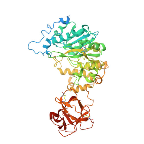
    6E7I, 6NQT

  • PubMed Abstract: 

    Studying posttranslational modifications classically relies on experimental strategies that oversimplify the complex biosynthetic machineries of living cells. Protein glycosylation contributes to essential biological processes, but correlating glycan structure, underlying protein, and disease-relevant biosynthetic regulation is currently elusive. Here, we engineer living cells to tag glycans with editable chemical functionalities while providing information on biosynthesis, physiological context, and glycan fine structure. We introduce a non-natural substrate biosynthetic pathway and use engineered glycosyltransferases to incorporate chemically tagged sugars into the cell surface glycome of the living cell. We apply the strategy to a particularly redundant yet disease-relevant human glycosyltransferase family, the polypeptide N-acetylgalactosaminyl transferases. This approach bestows a gain-of-chemical-functionality modification on cells, where the products of individual glycosyltransferases can be selectively characterized or manipulated to understand glycan contribution to major physiological processes.


  • Organizational Affiliation
    • Department of Chemistry, Stanford University, Stanford, CA 94305, USA; Chemical Glycobiology Laboratory, The Francis Crick Institute, NW1 1AT London, United Kingdom; Department of Chemistry, Imperial College London, W12 0BZ London, United Kingdom. Electronic address: b.schumann@imperial.ac.uk.

Macromolecule Content 

  • Total Structure Weight: 392.72 kDa 
  • Atom Count: 23,138 
  • Modeled Residue Count: 2,943 
  • Deposited Residue Count: 3,426 
  • Unique protein chains: 1

Macromolecules

Find similar proteins by:|  3D Structure
Entity ID: 1
MoleculeChains  Sequence LengthOrganismDetailsImage
Polypeptide N-acetylgalactosaminyltransferase 2
A, B, C, D, E
A, B, C, D, E, F
571Homo sapiensMutation(s): 2 
Gene Names: GALNT2
EC: 2.4.1.41
UniProt & NIH Common Fund Data Resources
Find proteins for Q10471 (Homo sapiens)
Explore Q10471 
Go to UniProtKB:  Q10471
PHAROS:  Q10471
GTEx:  ENSG00000143641 
Entity Groups
Sequence Clusters30% Identity50% Identity70% Identity90% Identity95% Identity100% Identity
UniProt GroupQ10471
Sequence Annotations
Expand
Reference Sequence

Small Molecules

Ligands 2 Unique
IDChains Name / Formula / InChI Key2D Diagram3D Interactions
LR7

Query on LR7


Download:Ideal Coordinates CCD File
H [auth A]
J [auth B]
L [auth C]
N [auth D]
P [auth E]
H [auth A],
J [auth B],
L [auth C],
N [auth D],
P [auth E],
R [auth F]
[[(2~{R},3~{S},4~{R},5~{R})-5-[2,4-bis(oxidanylidene)pyrimidin-1-yl]-3,4-bis(oxidanyl)oxolan-2-yl]methoxy-oxidanyl-phosphoryl] [(2~{R},3~{R},4~{R},5~{R},6~{R})-3-(hex-5-ynoylamino)-6-(hydroxymethyl)-4,5-bis(oxidanyl)oxan-2-yl] hydrogen phosphate
C21 H31 N3 O17 P2
SPUYDPWWABYYLH-SSBRDYMUSA-N
MN

Query on MN


Download:Ideal Coordinates CCD File
G [auth A]
I [auth B]
K [auth C]
M [auth D]
O [auth E]
G [auth A],
I [auth B],
K [auth C],
M [auth D],
O [auth E],
Q [auth F]
MANGANESE (II) ION
Mn
WAEMQWOKJMHJLA-UHFFFAOYSA-N

Experimental Data & Validation

Experimental Data

  • Method: X-RAY DIFFRACTION
  • Resolution: 3.05 Å
  • R-Value Free:  0.286 (Depositor) 
  • R-Value Work:  0.231 (Depositor) 
  • R-Value Observed: 0.234 (Depositor) 
Space Group: P 21 21 21
Unit Cell:
Length ( Å )Angle ( ˚ )
a = 116.575α = 90
b = 120.126β = 90
c = 247.389γ = 90
Software Package:
Software NamePurpose
REFMACrefinement
XDSdata reduction
SCALAdata scaling
PHASERphasing

Structure Validation

View Full Validation Report


Ligand Structure Quality Assessment 


Entry History 

Deposition Data

Revision History  (Full details and data files)

  • Version 1.0: 2020-01-29
    Type: Initial release
  • Version 1.1: 2020-08-12
    Changes: Database references, Derived calculations
  • Version 1.2: 2023-10-11
    Changes: Data collection, Database references, Refinement description
  • Version 1.3: 2024-10-23
    Changes: Structure summary