6NVQ | pdb_00006nvq

Crystal structure of the VASH1-SVBP complex


Experimental Data Snapshot

  • Method: X-RAY DIFFRACTION
  • Resolution: 2.10 Å
  • R-Value Free: 
    0.214 (Depositor), 0.230 (DCC) 
  • R-Value Work: 
    0.198 (Depositor), 0.206 (DCC) 
  • R-Value Observed: 
    0.198 (Depositor) 

wwPDB Validation 3D Report Full Report

Validation slider image for 6NVQ

This is version 1.3 of the entry. See complete history

Literature

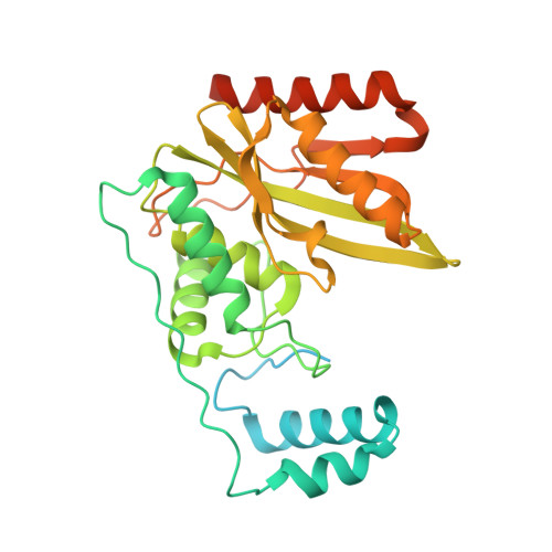
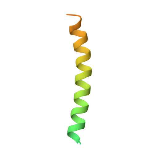
Crystal structure of the tubulin tyrosine carboxypeptidase complex VASH1-SVBP.

Adamopoulos, A.Landskron, L.Heidebrecht, T.Tsakou, F.Bleijerveld, O.B.Altelaar, M.Nieuwenhuis, J.Celie, P.H.N.Brummelkamp, T.R.Perrakis, A.

(2019) Nat Struct Mol Biol 26: 567-570

  • DOI: https://doi.org/10.1038/s41594-019-0254-6
  • Primary Citation Related Structures: 
    6NVQ

  • PubMed Abstract: 

    The cyclic enzymatic removal and ligation of the C-terminal tyrosine of α-tubulin generates heterogeneous microtubules and affects their functions. Here we describe the crystal and solution structure of the tubulin carboxypeptidase complex between vasohibin (VASH1) and small vasohibin-binding protein (SVBP), which folds in a long helix, which stabilizes the VASH1 catalytic domain. This structure, combined with molecular docking and mutagenesis experiments, reveals which residues are responsible for recognition and cleavage of the tubulin C-terminal tyrosine.


  • Organizational Affiliation
    • Division of Biochemistry, the Netherlands Cancer Institute, Amsterdam, the Netherlands.

Macromolecule Content 

  • Total Structure Weight: 44.44 kDa 
  • Atom Count: 2,322 
  • Modeled Residue Count: 272 
  • Deposited Residue Count: 391 
  • Unique protein chains: 2

Macromolecules

Find similar proteins by:|  3D Structure
Entity ID: 1
MoleculeChains  Sequence LengthOrganismDetailsImage
Tubulinyl-Tyr carboxypeptidase 1A [auth C]320Homo sapiensMutation(s): 1 
Gene Names: VASH1KIAA1036VASH
EC: 3.4.17.17
UniProt & NIH Common Fund Data Resources
Find proteins for Q7L8A9 (Homo sapiens)
Explore Q7L8A9 
Go to UniProtKB:  Q7L8A9
PHAROS:  Q7L8A9
GTEx:  ENSG00000071246 
Entity Groups
Sequence Clusters30% Identity50% Identity70% Identity90% Identity95% Identity100% Identity
UniProt GroupQ7L8A9
Sequence Annotations
Expand
Reference Sequence
Find similar proteins by:|  3D Structure
Entity ID: 2
MoleculeChains  Sequence LengthOrganismDetailsImage
Small vasohibin-binding protein71Homo sapiensMutation(s): 0 
Gene Names: SVBPCCDC23
UniProt & NIH Common Fund Data Resources
Find proteins for Q8N300 (Homo sapiens)
Explore Q8N300 
Go to UniProtKB:  Q8N300
PHAROS:  Q8N300
GTEx:  ENSG00000177868 
Entity Groups
Sequence Clusters30% Identity50% Identity70% Identity90% Identity95% Identity100% Identity
UniProt GroupQ8N300
Sequence Annotations
Expand
Reference Sequence

Experimental Data & Validation

Experimental Data

  • Method: X-RAY DIFFRACTION
  • Resolution: 2.10 Å
  • R-Value Free:  0.214 (Depositor), 0.230 (DCC) 
  • R-Value Work:  0.198 (Depositor), 0.206 (DCC) 
  • R-Value Observed: 0.198 (Depositor) 
Space Group: P 21 21 2
Unit Cell:
Length ( Å )Angle ( ˚ )
a = 70.453α = 90
b = 128.929β = 90
c = 45.222γ = 90
Software Package:
Software NamePurpose
REFMACrefinement
PDB_EXTRACTdata extraction
XDSdata reduction
SCALAdata scaling
CRANK2phasing

Structure Validation

View Full Validation Report


Entry History 

Deposition Data

Revision History  (Full details and data files)

  • Version 1.0: 2019-05-22
    Type: Initial release
  • Version 1.1: 2019-07-03
    Changes: Data collection, Database references
  • Version 1.2: 2019-07-17
    Changes: Data collection, Database references
  • Version 1.3: 2024-05-15
    Changes: Data collection, Database references