6O0P

crystal structure of BCL-2 G101A mutation with venetoclax


Experimental Data Snapshot

  • Method: X-RAY DIFFRACTION
  • Resolution: 1.80 Å
  • R-Value Free: 0.232 
  • R-Value Work: 0.193 
  • R-Value Observed: 0.195 

wwPDB Validation   3D Report Full Report

Currently 6O0P does not have a validation slider image.


This is version 2.2 of the entry. See complete history


Literature

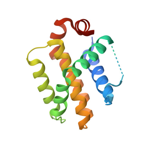
Structures of BCL-2 in complex with venetoclax reveal the molecular basis of resistance mutations.

Birkinshaw, R.W.Gong, J.N.Luo, C.S.Lio, D.White, C.A.Anderson, M.A.Blombery, P.Lessene, G.Majewski, I.J.Thijssen, R.Roberts, A.W.Huang, D.C.S.Colman, P.M.Czabotar, P.E.

(2019) Nat Commun 10: 2385-2385

  • DOI: https://doi.org/10.1038/s41467-019-10363-1
  • Primary Citation of Related Structures:  
    6O0K, 6O0L, 6O0M, 6O0O, 6O0P

  • PubMed Abstract: 

    Venetoclax is a first-in-class cancer therapy that interacts with the cellular apoptotic machinery promoting apoptosis. Treatment of patients suffering chronic lymphocytic leukaemia with this BCL-2 antagonist has revealed emergence of a drug-selected BCL-2 mutation (G101V) in some patients failing therapy. To understand the molecular basis of this acquired resistance we describe the crystal structures of venetoclax bound to both BCL-2 and the G101V mutant. The pose of venetoclax in its binding site on BCL-2 reveals small but unexpected differences as compared to published structures of complexes with venetoclax analogues. The G101V mutant complex structure and mutant binding assays reveal that resistance is acquired by a knock-on effect of V101 on an adjacent residue, E152, with venetoclax binding restored by a E152A mutation. This provides a framework for considering analogues of venetoclax that might be effective in combating this mutation.


  • Organizational Affiliation

    Walter and Eliza Hall Institute of Medical Research, 1G Royal Parade, Parkville, VIC, 3052, Australia. birkinshaw.r@wehi.edu.au.


Macromolecules
Find similar proteins by:  (by identity cutoff)  |  3D Structure
Entity ID: 1
MoleculeChains Sequence LengthOrganismDetailsImage
Apoptosis regulator Bcl-2,Bcl-2-like protein 1,Apoptosis regulator Bcl-2166Homo sapiensMutation(s): 1 
Gene Names: BCL2BCL2L1BCL2LBCLX
UniProt & NIH Common Fund Data Resources
Find proteins for Q07817 (Homo sapiens)
Explore Q07817 
Go to UniProtKB:  Q07817
PHAROS:  Q07817
GTEx:  ENSG00000171552 
Find proteins for P10415 (Homo sapiens)
Explore P10415 
Go to UniProtKB:  P10415
PHAROS:  P10415
GTEx:  ENSG00000171791 
Entity Groups  
Sequence Clusters30% Identity50% Identity70% Identity90% Identity95% Identity100% Identity
UniProt GroupsQ07817P10415
Sequence Annotations
Expand
  • Reference Sequence
Small Molecules
Ligands 2 Unique
IDChains Name / Formula / InChI Key2D Diagram3D Interactions
LBM
Query on LBM

Download Ideal Coordinates CCD File 
B [auth A]4-{4-[(4'-chloro-5,5-dimethyl[3,4,5,6-tetrahydro[1,1'-biphenyl]]-2-yl)methyl]piperazin-1-yl}-N-[(3-nitro-4-{[(oxan-4-yl )methyl]amino}phenyl)sulfonyl]-2-[(1H-pyrrolo[2,3-b]pyridin-5-yl)oxy]benzamide
C45 H50 Cl N7 O7 S
LQBVNQSMGBZMKD-UHFFFAOYSA-N
PEG
Query on PEG

Download Ideal Coordinates CCD File 
C [auth A],
D [auth A],
E [auth A]
DI(HYDROXYETHYL)ETHER
C4 H10 O3
MTHSVFCYNBDYFN-UHFFFAOYSA-N
Binding Affinity Annotations 
IDSourceBinding Affinity
LBM Binding MOAD:  6O0P Ki: 0.11 (nM) from 1 assay(s)
Experimental Data & Validation

Experimental Data

  • Method: X-RAY DIFFRACTION
  • Resolution: 1.80 Å
  • R-Value Free: 0.232 
  • R-Value Work: 0.193 
  • R-Value Observed: 0.195 
  • Space Group: P 21 21 21
Unit Cell:
Length ( Å )Angle ( ˚ )
a = 33.398α = 90
b = 48.945β = 90
c = 86.474γ = 90
Software Package:
Software NamePurpose
PHENIXrefinement
XDSdata reduction
XDSdata scaling
PHASERphasing

Structure Validation

View Full Validation Report

Currently 6O0P does not have a validation slider image.


Entry History 

Deposition Data

Revision History  (Full details and data files)

  • Version 1.0: 2019-05-22
    Type: Initial release
  • Version 1.1: 2019-06-19
    Changes: Data collection, Database references
  • Version 1.2: 2019-07-10
    Changes: Data collection, Database references, Source and taxonomy, Structure summary
  • Version 2.0: 2019-11-20
    Changes: Derived calculations, Non-polymer description, Structure summary
  • Version 2.1: 2019-12-11
    Changes: Structure summary
  • Version 2.2: 2023-10-11
    Changes: Data collection, Database references, Derived calculations, Refinement description, Structure summary